Neuromelanin, the enigmatic dark pigment that accumulates in specific brain regions throughout our lives, represents one of the most intriguing yet understudied aspects of human neurobiology. Unlike its skin-protecting cousin, this brain-specific melanin may hold keys to understanding both cognitive vitality and neurodegenerative disease.
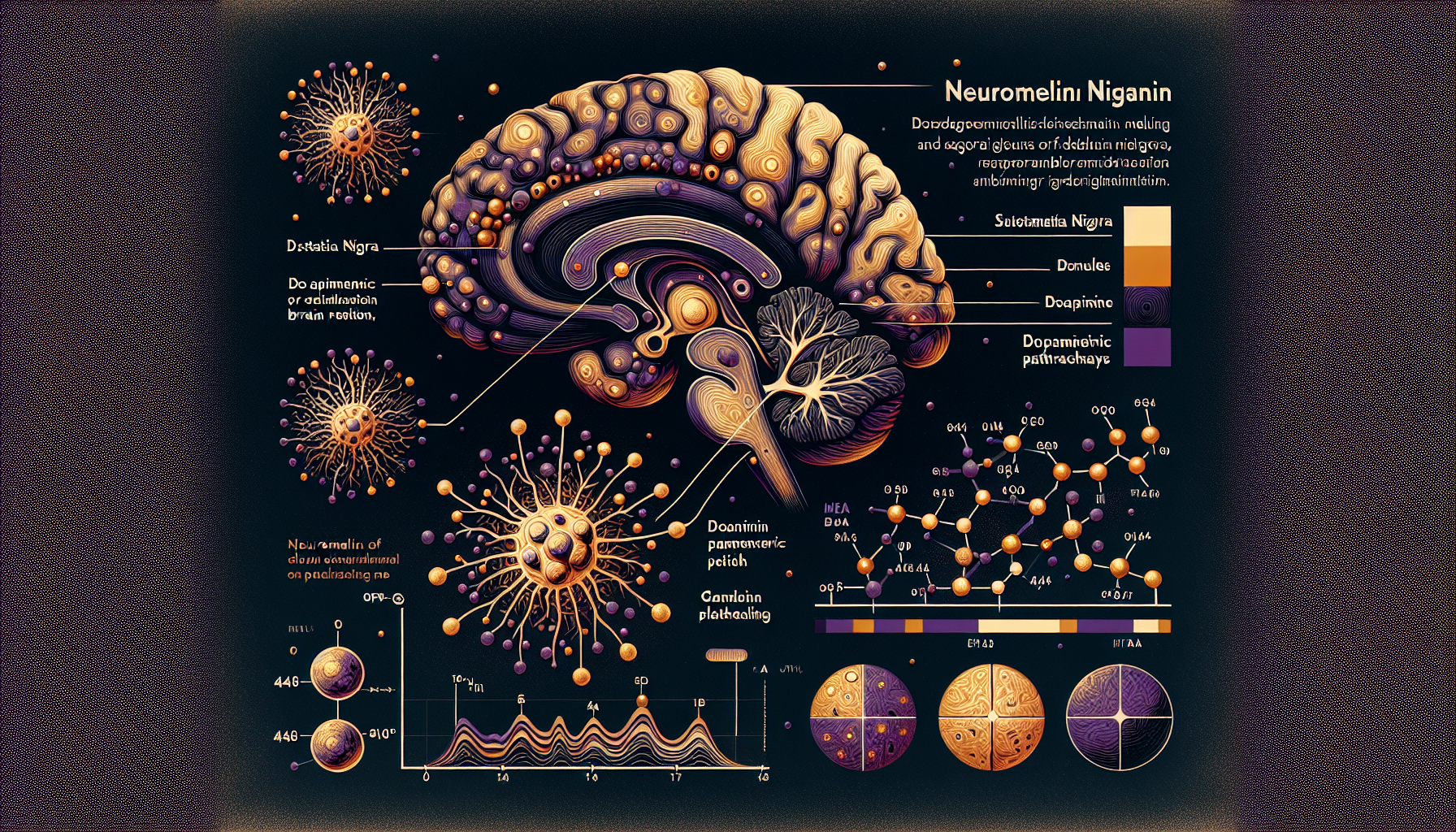
In the depths of the human brainstem, within clusters of neurons that control movement and arousal, a remarkable process unfolds over decades. Dopamine-producing cells in the substantia nigra and norepinephrine neurons in the locus coeruleus gradually accumulate dark granules of neuromelanin—a complex polymer that appears nowhere else in the body with such specificity and abundance. This pigment, virtually absent at birth, steadily increases throughout life, creating the characteristic dark coloration that gives the substantia nigra its name.
Yet this accumulation presents a paradox that has puzzled neuroscientists for generations. The very neurons that produce the most neuromelanin are precisely those most vulnerable in Parkinson's disease, Alzheimer's disease, and other age-related neurodegenerative conditions. Is neuromelanin a protective guardian, a toxic burden, or something more complex?
The Chemistry of Neural Protection
Neuromelanin differs fundamentally from the eumelanin found in skin and hair. While both share the ability to bind metals and neutralize free radicals, neuromelanin incorporates unique elements that reflect its specialized neural environment. Research by Zecca and colleagues at the Italian National Research Council has revealed that neuromelanin forms through the oxidation of dopamine and norepinephrine—the very neurotransmitters these neurons produce and release.
This formation process appears to serve as a cellular cleanup mechanism. When dopamine metabolism produces potentially harmful byproducts like dopamine quinones and reactive oxygen species, neuromelanin synthesis provides a pathway to sequester these toxic intermediates. The pigment acts like molecular flypaper, binding not only these organic toxins but also metal ions, particularly iron and copper, which can catalyze destructive oxidative reactions.
Studies using electron paramagnetic resonance (EPR) spectroscopy have shown that neuromelanin contains stable free radicals similar to those found in other melanins, but with distinct characteristics that suggest specialized antioxidant properties. The pigment's ability to chelate iron is particularly crucial, as iron accumulation in the brain correlates strongly with neurodegeneration and cognitive decline.
The Parkinson's Paradox
The relationship between neuromelanin and Parkinson's disease reveals the complexity of this pigment's role in brain health. Post-mortem studies consistently show that Parkinson's patients have dramatically reduced neuromelanin levels in the substantia nigra—not because the pigment causes disease, but because the neurons that produce it are dying.
However, the story is more nuanced than simple cell death. Research by Sulzer and colleagues at Columbia University has demonstrated that neuromelanin can become immunogenic under certain conditions. When neurons die and release their neuromelanin granules, the pigment can activate microglia—the brain's immune cells—potentially triggering inflammatory responses that accelerate neurodegeneration.
This discovery has led to the "neuromelanin hypothesis" of Parkinson's disease progression. Early in the disease process, neuromelanin may indeed be protective, helping neurons cope with oxidative stress and toxic metal accumulation. But as neurons become overwhelmed and begin to die, the release of neuromelanin granules may transform this protective molecule into an inflammatory signal, creating a vicious cycle of neurodegeneration.
The timing appears critical. Young, healthy dopamine neurons with moderate neuromelanin levels show enhanced resistance to oxidative stress. But aged neurons with heavy neuromelanin loads may reach a tipping point where the pigment's protective capacity is exceeded by its potential for inflammatory activation.
Beyond Movement: Cognitive Connections
While neuromelanin's role in Parkinson's disease has received the most attention, emerging evidence suggests its influence extends far beyond motor control. The locus coeruleus, which contains the brain's highest concentration of neuromelanin-producing neurons, serves as the primary source of norepinephrine throughout the brain—a neurotransmitter crucial for attention, arousal, and cognitive flexibility.
Recent neuroimaging studies have begun to reveal correlations between locus coeruleus neuromelanin levels and cognitive performance in healthy aging. Using specialized MRI techniques that can detect neuromelanin's unique magnetic properties, researchers have found that individuals with higher locus coeruleus neuromelanin show better performance on tasks requiring sustained attention and working memory.
This finding suggests that neuromelanin accumulation, rather than being purely pathological, may serve as a biomarker of neuronal health and resilience. Neurons capable of maintaining high neuromelanin levels throughout aging may be those best equipped to handle the metabolic demands of neurotransmitter synthesis and the oxidative stress of normal brain function.
The Iron Connection
Perhaps nowhere is neuromelanin's protective role more evident than in iron homeostasis. The human brain accumulates iron throughout life, and excessive iron deposition is implicated in multiple neurodegenerative diseases. Neuromelanin's ability to chelate iron provides a mechanism for neurons to manage this potentially toxic metal.
Studies using synchrotron X-ray techniques have revealed that neuromelanin granules contain remarkably high concentrations of iron—up to 180 times higher than surrounding tissue. Rather than representing pathological iron overload, this appears to be protective sequestration. The iron bound to neuromelanin is largely in the less reactive Fe3+ form and is prevented from participating in the Fenton reaction that generates destructive hydroxyl radicals.
This iron-binding capacity may explain why dopamine neurons, despite their high metabolic activity and oxidative stress exposure, can survive for decades. The gradual accumulation of neuromelanin provides an expanding capacity for iron detoxification, potentially explaining why neurodegenerative diseases typically emerge in later life when this protective system becomes overwhelmed.
Key Takeaways
• Neuromelanin forms through the oxidation of dopamine and norepinephrine, serving as a cellular detoxification system that sequesters toxic metabolites and metal ions throughout life.
• The pigment's protective effects appear to be dose-dependent and age-related, providing neuroprotection in healthy neurons but potentially contributing to inflammation when released from dying cells.
• Neuromelanin loss in the substantia nigra serves as both a consequence and potential accelerator of Parkinson's disease progression through inflammatory mechanisms.
• Higher neuromelanin levels in the locus coeruleus correlate with better cognitive performance in healthy aging, suggesting the pigment may serve as a biomarker of neuronal resilience.
• The pigment's iron-chelating properties provide a crucial mechanism for managing brain iron accumulation, which increases throughout life and contributes to neurodegeneration when dysregulated.
• Understanding neuromelanin's dual nature—protective in health, inflammatory in disease—may reveal new therapeutic targets for age-related neurodegeneration.
References
Zecca, L., Youdim, M. B., Riederer, P., Connor, J. R., & Crichton, R. R. "Iron, brain ageing and neurodegenerative disorders." Nature Reviews Neuroscience 5(11), 863-873 (2004).
Sulzer, D., Cassidy, C., Horga, G., Kang, U. J., Fahn, S., Casella, L., ... & Zecca, L. "Neuromelanin detection by magnetic resonance imaging (MRI) and its promise as a biomarker for Parkinson's disease." npj Parkinson's Disease 4(1), 1-13 (2018).
Zucca, F. A., Segura-Aguilar, J., Ferrari, E., Muñoz, P., Paris, I., Sulzer, D., ... & Zecca, L. "Interactions of iron, dopamine and neuromelanin pathways in brain aging and Parkinson's disease." Progress in Neurobiology 155, 96-119 (2017).
Cassidy, C. M., & Zucca, F. A. "Neuromelanin, neuromelanin-sensitive MRI, and noradrenergic dysfunction in Parkinson's disease." Journal of Neural Transmission 126(11), 1433-1449 (2019).
McGeer, P. L., & McGeer, E. G. "Glial reactions in Parkinson's disease." Movement Disorders 23(4), 474-483 (2008).
Fedorow, H., Tribl, F., Halliday, G., Gerlach, M., Riederer, P., & Double, K. L. "Neuromelanin in human dopamine neurons: comparison with peripheral melanins and relevance to Parkinson's disease." Progress in Neurobiology 75(2), 109-124 (2005).