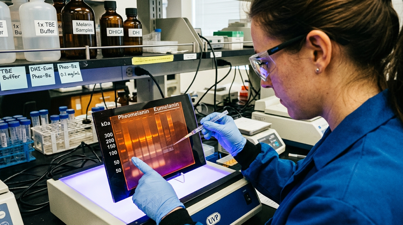
The designation "melanin" is often used as a monolith, a single dark pigment responsible for coloration and sun protection. Yet, this simplification obscures a critical biological reality: melanin is not one substance, but a family of heterogeneous polymers with dramatically different—and in some cases, opposing—functions. This is most starkly illustrated by the case of individuals with red hair and fair skin, who possess an abundance of melanin, yet experience a significantly higher risk for UV-induced skin cancers. The paradox resolves when we understand that their melanosomes are rich in the sulfur-containing pheomelanin, a molecule whose biophysical response to light is fundamentally different from its more common cousin, eumelanin.
This divergence is not merely a chemical curiosity; it is a distinction with profound biophysical consequences that extend from photoprotection and oxidative stress to the frontiers of bioelectric signaling. The ratio of eumelanin to pheomelanin in a tissue dictates a complex interplay of energy absorption, dissipation, and transfer. Understanding the architectural differences between these two polymers is therefore essential to appreciating melanin’s true role as an active, tunable biophysical agent within the cellular environment, rather than a passive pigment.
The Architectural Blueprint: From Monomers to Macromolecular Function
The functional schism between eumelanin and pheomelanin begins at the level of their monomeric building blocks. Both are synthesized from the amino acid tyrosine, but a key branching point in the melanogenesis pathway determines their ultimate structure and properties.
Eumelanin is a nitrogen-based polymer primarily composed of 5,6-dihydroxyindole (DHI) and 5,6-dihydroxyindole-2-carboxylic acid (DHICA). These planar, indole-based molecules polymerize into what is often described as a disordered, stacked aggregate. Imagine small, graphite-like sheets layered imperfectly upon one another, creating a complex, three-dimensional macromolecule. This structural disorder is not a defect but a feature. It creates a vast landscape of electronic energy levels, which is the physical basis for eumelanin's most famous characteristic: its ability to absorb a remarkably broad spectrum of electromagnetic radiation, from UV-C (<280 nm) through the visible and into the near-infrared. The DHI/DHICA ratio itself can fine-tune these properties, with DHI-rich polymers exhibiting different redox behavior than their DHICA-rich counterparts.
Pheomelanin, by contrast, incorporates sulfur into its structure. Its synthesis involves the reaction of dopaquinone (an intermediate in the melanin pathway) with the sulfur-containing amino acid cysteine. This leads to the formation of cysteinyldopas, which then polymerize into macromolecules characterized by benzothiazine and benzothiazole units. The presence of the sulfur atom is the critical architectural change. It disrupts the stable, stacked indole structure of eumelanin, creating what is often referred to as a more open and structurally flexible polymer. This sulfur linkage is, from a chemical standpoint, a point of vulnerability—a "weak link" that fundamentally alters how the molecule interacts with and processes energy.
Energy Handling: Photoprotective Sink vs. Phototoxic Source
When a photon of UV radiation strikes a melanin polymer, its fate is determined by the architecture described above. This is where the functional divergence becomes a matter of cellular life and death.
Eumelanin is arguably one of the most effective photoprotective materials in the natural world. Research by groups like those of Patrick Meredith and Tadeusz Sarna has shown that upon absorbing a photon, the eumelanin polymer undergoes an astonishingly rapid process of ultrafast internal conversion. More than 99.9% of the absorbed photon energy is dissipated non-radiatively as heat on a femtosecond-to-picosecond timescale. The energy is shunted through the polymer's complex electronic landscape and released harmlessly before it has a chance to generate damaging chemical species. Eumelanin acts as the ultimate energy sink, a molecular "black hole" for high-energy photons.
Pheomelanin behaves in a tragically different manner. When it absorbs UV energy, the weaker bonds associated with its sulfur-containing benzothiazine units make it susceptible to photochemical degradation. Instead of harmlessly dissipating the energy as heat, a significant fraction can be transferred to molecular oxygen, generating highly destructive reactive oxygen species (ROS), such as the superoxide anion and singlet oxygen. Pheomelanin, therefore, acts as an endogenous photosensitizer. It takes the sun's energy and weaponizes it against the cell. This mechanism, demonstrated in studies by researchers like Douglas Brash at Yale, is a primary driver for the elevated risk of melanoma in individuals with high pheomelanin content. The very molecule evolved for pigmentation becomes a source of carcinogenic stress.
The Redox Divide: Stable Radicals and Bioelectric Influence
The biophysical differences extend far beyond photochemistry into the realm of electrochemistry and redox biology. Both melanins are redox-active, meaning they can readily accept and donate electrons, but they do so with opposite net effects on the cellular environment.
Eumelanin is characterized by its nature as a stable free radical, a property easily detectable using Electron Paramagnetic Resonance (EPR) spectroscopy. This intrinsic radical population allows it to act as a powerful antioxidant, scavenging and neutralizing other, more damaging free radicals. It functions as a redox buffer, absorbing oxidative insults. Furthermore, eumelanin exhibits semiconductor properties. Landmark work by John McGinness and colleagues in the 1970s first proposed melanin as an amorphous semiconductor, capable of switching between high- and low-conductivity states. Its conductivity is highly dependent on hydration, and it can conduct both electrons and protons, suggesting a potential role in local bioelectric circuits. This ability to manage and buffer charge could influence the membrane potential (Vmem) of nearby cells, a critical parameter in controlling cell proliferation, differentiation, and migration, as shown by the work of Michael Levin's group at Tufts University.
Pheomelanin, again, presents a stark contrast. Its redox cycling tends to deplete the cell's primary antioxidant, glutathione, during its synthesis and subsequent reactions. Instead of buffering against oxidative stress, it can be a source of it, even in the absence of light. Studies have shown that pheomelanin can induce DNA damage through ROS generation in a process known as chemiexcitation. From a bioelectric standpoint, a polymer that actively generates ROS and disrupts the local redox balance is likely to have a destabilizing effect on cellular membrane potentials, potentially contributing to the aberrant signaling observed in cancer. The two polymers thus represent opposing poles in the maintenance of cellular homeostasis: eumelanin the stabilizer, pheomelanin the destabilizer.
Key Takeaways
- The biophysical functions of melanin are determined by its chemical structure, with eumelanin being an indole-based polymer and pheomelanin a sulfur-containing benzothiazine polymer.
- Eumelanin provides exceptional photoprotection by converting over 99.9% of absorbed UV radiation into heat through ultrafast internal conversion, acting as an energy sink.
- Pheomelanin acts as an endogenous photosensitizer, generating damaging reactive oxygen species (ROS) upon UV exposure, which directly contributes to a higher risk of melanoma.
- The ratio of eumelanin to pheomelanin, not just the total melanin concentration, is a more accurate predictor of an individual's susceptibility to sun-induced skin damage and cancer.
- Beyond photochemistry, eumelanin is a stable free radical and redox buffer that can conduct charge, while pheomelanin can promote oxidative stress, suggesting these polymers have opposing influences on cellular bioelectric states.
- These fundamental differences underscore the need to move beyond viewing melanin as a simple pigment and to investigate its active role as a complex, tunable biopolymer in physiology and pathology.
References
- d'Ischia, M., Wakamatsu, K., Cicoira, F., Di Mauro, E., Garcia-Borron, J. C., Commo, S., Galván, I., Ghanem, G., Kenzo, K., Meredith, P., Pezzella, A., Santato, C., Sarna, T., Simon, J. D., & Ito, S. "Melanins and melanogenesis: from pigment cells to human health and technological applications." Pigment Cell & Melanoma Research 28(5), 520-544 (2015). DOI: 10.1111/pcmr.12393
- Mitra, D., Luo, X., Morgan, A., Wang, J., Hoang, M. P., Lo, J., Guerrero, C. R., Lennerz, J. K., Mihm, M. C., Wargo, J. A., Robinson, K. C., Devereux, W. L., Tang, C., Fisher, D. E. "An ultraviolet-radiation-independent pathway to melanoma carcinogenesis in the red hair/fair skin background." Nature 491(7424), 449-453 (2012). DOI: 10.1038/nature11624
- Meredith, P., & Sarna, T. "The physical and chemical properties of eumelanin." Pigment Cell Research 19(6), 572-594 (2006). DOI: 10.1111/j.1600-0749.2006.00345.x
- McGinness, J., Corry, P., & Proctor, P. "Amorphous semiconductor switching in melanins." Science 183(4127), 853-855 (1974). DOI: 10.1126/science.183.4127.853
- Pezzella, A., Panzella, L., Riccio, G., d'Ischia, M., & Napolitano, A. "The truth, the whole truth, and nothing but the truth about pheomelanin." International Journal of Molecular Sciences 23(16), 9037 (2022). DOI: 10.3390/ijms23169037
- Simon, J. D., & Peles, D. "The red-head's dilemma: the genetics and chemistry of pheomelanin." Accounts of Chemical Research 42(4), 543-550 (2009). DOI: 10.1021/ar800168p
- Zecca, L., Fariello, R., Riederer, P., Sulzer, D., Gatti, A., & Tampellini, D. "The absolute concentration of human neuromelanin: a postmortem analysis." Journal of Neurochemistry 98(3), 857-865 (2006). DOI: 10.1111/j.1471-4159.2006.03932.x
- Levin, M. "Bioelectric signaling: Reprogrammable circuits underlying embryonic development, regeneration, and cancer." Cell 184(8), 1971-1989 (2021). DOI: 10.1016/j.cell.2021.02.034