Deep within the human brainstem, a specialized form of melanin silently accumulates over the course of our lifetimes, turning specific neural populations a deep, distinct black. Once dismissed as a mere metabolic waste product, neuromelanin is now recognized as a highly active biophysical sink and iron chelator that directly dictates the survival or death of our most vulnerable neurons.
When 19th-century neuroanatomists first sliced into the human brainstem, they were struck by two dark, wing-like structures: the substantia nigra (black substance) and the locus coeruleus (blue spot). Unlike the melanin in our skin, which is synthesized early in life to protect against ultraviolet radiation, this deep-brain pigment is entirely absent at birth. It accumulates linearly with age, darkening year by year, exclusively in the neurons responsible for producing our most critical catecholamine neurotransmitters: dopamine and norepinephrine.
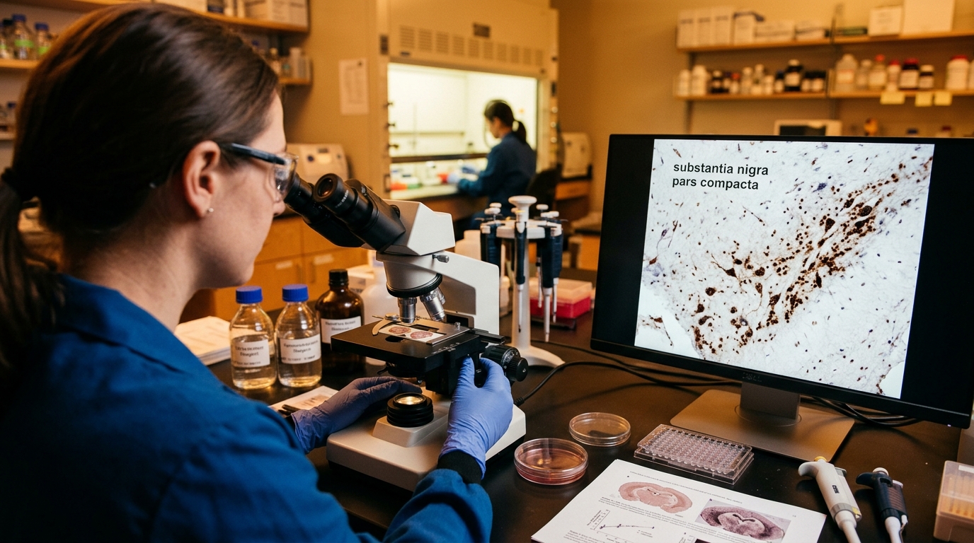
For decades, the presence of this pigment was treated as a biological curiosity. However, the exact regions where this pigment is most dense are also the epicenters of neurodegenerative collapse. In Parkinson’s disease, the dark streaks of the substantia nigra physically fade as pigmented neurons undergo massive cell death. This visually striking pathology raises a provocative question: does this pigment evolve to protect our neural circuitry, or does it eventually become the very agent of its destruction? Modern biophysics and molecular neurology are finally converging on an answer, revealing neuromelanin as an extraordinary biological capacitor that walks a knife-edge between neuroprotection and neurotoxicity.
The Architecture of a Cellular Sponge
To understand neuromelanin’s function, one must first understand its unconventional origins. In the periphery of the body, melanin is synthesized via specialized enzymes like tyrosinase. In the brain, however, neuromelanin is largely born out of biochemical necessity and oxidative hazard.
Dopamine is a highly reactive molecule. When it is properly sequestered inside synaptic vesicles, it is safe. But cytosolic dopamine—the dopamine floating freely within the main body of the neuron—is exceptionally dangerous. If left unchecked, it spontaneously oxidizes into toxic quinones that can shred cellular machinery and induce oxidative stress. Work pioneered by researchers like David Sulzer at Columbia University and Luigi Zecca at the Italian National Research Council (CNR) demonstrated that neuromelanin synthesis is driven by the autoxidation of this excess cytosolic dopamine.
Rather than letting toxic dopamine metabolites destroy the cell, the neuron polymerizes them into a dense, insoluble matrix. The resulting structure is highly complex. Biophysical analyses, such as those conducted by Simon and colleagues using photoelectron emission microscopy, reveal that neuromelanin possesses a spherical architecture featuring a pheomelanin-like core (rich in sulfur and benzothiazine units) surrounded by a highly protective eumelanin-like surface (derived from indole quinones). This melanic shell envelops a core of lipids and degraded proteins. In a biophysical sense, neuromelanin acts as a molecular sponge, safely locking away reactive metabolic byproducts that would otherwise trigger apoptotic cell death.
Iron Chelation and the Neuroprotective Mandate
Neuromelanin’s protective role extends far beyond trapping errant dopamine; it is the brain’s premier manager of heavy metals. The substantia nigra naturally contains some of the highest concentrations of iron in the human body. Iron is an absolute requirement for the enzyme tyrosine hydroxylase, which synthesizes dopamine. However, free, redox-active iron is highly volatile. Through the Fenton reaction, free iron catalyzes the production of highly destructive hydroxyl radicals.
Neuromelanin functions as an extraordinarily high-capacity iron chelator. It binds transition metals, effectively disarming them. The polymer structure of melanin features millions of binding sites capable of trapping $Fe^{3+}$ and holding it in a redox-silent state. Because of its unique biophysical properties—including its stable free radicals, measurable via Electron Paramagnetic Resonance (EPR) spectroscopy, and its broadband absorption—neuromelanin can safely dissipate the oxidative energy generated by these trapped metals without degrading.
For decades of a human lifespan, this system works flawlessly. The expanding volume of neuromelanin in our midbrain acts as a protective reservoir, keeping the cytosol free of both toxic dopamine quinones and rogue iron molecules, thereby ensuring the longevity of our critical motor and cognitive control networks.
The Tipping Point: From Shield to Neurotoxin
If neuromelanin is so protective, why is its presence the defining vulnerability in Parkinson’s disease? The answer lies in the physics of saturation and cellular capacitance.
As humans age, the neuromelanin polymer continuously binds iron and other metals, including copper and zinc. Eventually, the structural binding sites on the melanin matrix reach their maximum capacity. At this saturation point, the pigment can no longer act as a safe chelator. Instead, the heavily metallized surface of the neuromelanin molecule itself becomes a generator of reactive oxygen species.
When a heavily pigmented, overburdened neuron finally dies, it ruptures, spilling its massive payload of insoluble, iron-rich neuromelanin into the extracellular space. This is where the biological cascade turns catastrophic. Extracellular neuromelanin is highly immunogenic. When the brain's resident immune cells, microglia, encounter this dense, metal-studded biopolymer, they recognize it as a foreign body. They engulf the neuromelanin and release pro-inflammatory cytokines, triggering localized neuroinflammation.
This creates a self-propagating cycle of destruction: a dying neuron releases neuromelanin; the released neuromelanin activates microglia; the inflamed microglia release factors that kill neighboring dopamine neurons; those dying neurons release more neuromelanin. This mechanism elegantly explains the progressive, localized nature of substantia nigra degeneration in Parkinson's disease.
The Bioelectric Horizon: Viewing Pigment as a Semiconductor
While classical neuroscience views neuromelanin primarily through a biochemical lens, emerging biophysical frameworks suggest a much more profound role for this pigment. The Quantum Melanin Research Foundation advocates for the integration of solid-state physics and bioelectricity into our understanding of neural pigment.
Melanin is known to exhibit semiconductor-like behavior, with a defined bandgap (approximately ~1.85 eV for eumelanin), hydration-dependent proton conductivity, and robust electron-transfer capabilities. We must ask: what does the presence of a dense, semiconducting biopolymer do to the local electrical environment of a neuron?
Drawing on the theoretical frameworks of ion-channel computation and bioelectric morphology—such as the membrane potential ($V_{mem}$) signaling paradigms explored by the Levin lab at Tufts University—it is entirely plausible that neuromelanin influences the electrochemical gradients of the substantia nigra. A massive intracellular matrix of proton-conducting, electron-dense melanin likely alters a neuron's capacitance and its handling of local charge. When Parkinson’s disease strips the substantia nigra of this pigment, it may not just be a loss of a chemical buffer, but a fundamental alteration of the tissue’s bioelectric architecture and quantum tunneling environments.
Future therapeutic interventions may pivot away from simply trying to replace lost dopamine. Instead, they might focus on "desaturating" existing neuromelanin using targeted chelators (such as deferiprone) to restore its protective capacitance, or employing neuromodulation techniques designed to interact specifically with the biophysical properties of the melanin matrix. By understanding neuromelanin not as inert cellular debris, but as a dynamic, conductive bio-sponge, we open entirely new frontiers in the preservation of cognitive function and the treatment of neurodegenerative disease.
Key Takeaways
- Distinct Origin: Unlike the enzyme-driven synthesis of melanin in the skin, neuromelanin is formed primarily through the autoxidation of excess cytosolic dopamine, creating a safe storage mechanism for toxic metabolic byproducts.
- Structural Complexity: Neuromelanin exhibits a spherical architecture, utilizing a protective eumelanin-like surface to encapsulate a pheomelanin-like core, lipids, and degraded proteins.
- Vital Iron Chelation: The pigment serves a critical neuroprotective function by binding free iron, preventing the Fenton reaction, and trapping reactive oxygen species within its stable free radical matrix.
- The Saturation Threshold: As neurons age, neuromelanin can reach its maximum metal-binding capacity; at this point, it transitions from a protective antioxidant to a pro-oxidant liability.
- Driver of Neuroinflammation: When a dopamine neuron dies and ruptures, it dumps insoluble neuromelanin into the extracellular space, triggering severe microglial activation and the chronic neuroinflammation characteristic of Parkinson’s disease.
- Biophysical Implications: Beyond biochemistry, neuromelanin’s properties as an organic semiconductor and proton conductor suggest it may play an unrecognized role in modulating the local bioelectric and capacitive environment of midbrain neurons.
References
- Zecca, L., Tampellini, D., Gerlach, M., Riederer, P., Fariello, R. G., & Sulzer, D. "Substantia nigra neuromelanin: structure, synthesis, and molecular behaviour." Molecular Pathology 54(6), 414–418 (2001). DOI: 10.1136/mp.54.6.414.
- Sulzer, D., Bogulavsky, J., Larsen, K. E., Behr, G., Karatekin, E., Kleinman, M. H., Turro, N., Krantz, D., Edwards, R. H., Greene, L. A., & Zecca, L. "Neuromelanin biosynthesis is driven by excess cytosolic catecholamines not accumulated by synaptic vesicles." Proceedings of the National Academy of Sciences 97(22), 11869-11874 (2000). DOI: 10.1073/pnas.97.22.11869.
- Zucca, F. A., Segura-Aguilar, J., Ferrari, E., Muñoz, P., Paris, I., Sulzer, D., Sarna, T., Casella, L., & Zecca, L. "Interactions of iron, dopamine and neuromelanin pathways in brain aging and Parkinson's disease." Progress in Neurobiology 155, 96-119 (2017). DOI: 10.1016/j.pneurobio.2015.09.012.
- Bush, W. D., Garguilo, J., Zucca, F. A., Albertini, A., Zecca, L., Edwards, G. S., & Simon, J. D. "The surface oxidation potential of human neuromelanin reveals a spherical architecture with a pheomelanin core and a eumelanin surface." Proceedings of the National Academy of Sciences 103(40), 14785-14789 (2006). DOI: 10.1073/pnas.0605073103.
- Double, K. L., Zecca, L., Costi, P., Mauer, M., Griesinger, C., Ito, S., Ben-Shachar, D., Bringmann, G., Fariello, R. G., Riederer, P., & Gerlach, M. "Structural characteristics of human substantia nigra neuromelanin and synthetic dopamine melanins." Journal of Neurochemistry 75(6), 2583-2589 (2000). DOI: 10.1046/j.1471-4159.2000.0752583.x.
- Carballo-Carbajal, I., Francardo, V., Siret, C., et al. "Brain tyrosinase overexpression implicates age-dependent neuromelanin production in Parkinson’s disease pathogenesis." Nature Communications 10, 973 (2019). DOI: 10.1038/s41467-019-08858-y.