Current skin classification systems fail to capture melanin's complex biophysical properties, limiting both clinical precision and our understanding of this remarkable biomaterial's role in human physiology. A new framework based on melanin's electrical, optical, and chemical characteristics could revolutionize personalized medicine and unlock deeper insights into skin biology.
When dermatologist Thomas Fitzpatrick introduced his six-category skin typing system in 1975, he solved an immediate clinical problem: predicting how different individuals would respond to UV radiation. But nearly five decades later, this binary approach to melanin—essentially "how much" rather than "what kind"—has become a scientific bottleneck. Recent advances in melanin biophysics reveal that this ancient biomolecule functions as far more than a passive UV filter. It operates as a bioelectric semiconductor, a proton conductor, and potentially even a quantum information processor. These discoveries demand a classification system that matches melanin's true complexity.
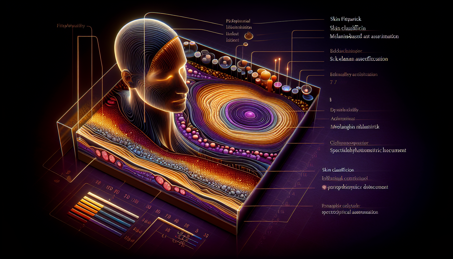
The Fitzpatrick Limitation: Missing the Biophysical Forest for the Pigmentation Trees
The Fitzpatrick scale's six types, ranging from "always burns, never tans" (Type I) to "never burns, always tans" (Type VI), capture only melanin's photoprotective capacity. This approach overlooks critical biophysical variations that exist even within the same Fitzpatrick category. Two individuals classified as Type III, for instance, may have dramatically different ratios of eumelanin (the brown-black, photoprotective polymer) to pheomelanin (the red-yellow, potentially photosensitizing variant). This ratio affects not just UV response, but also electrical conductivity, free radical scavenging capacity, and metal chelation properties.
Research by Ito and Wakamatsu at Fujita Health University has demonstrated that eumelanin-to-pheomelanin ratios vary dramatically across populations and even within families, independent of visible pigmentation levels. Their alkaline hydrogen peroxide oxidation method reveals that some fair-skinned individuals possess higher eumelanin ratios than their darker-skinned counterparts—a finding that challenges fundamental assumptions about melanin distribution and function.
The clinical implications extend beyond dermatology. Neuromelanin in the substantia nigra, chemically distinct from skin melanin, accumulates iron and may play crucial roles in Parkinson's disease pathogenesis. Yet current research often extrapolates from skin pigmentation to neurological melanin function—a leap that ignores the unique biophysical properties of different melanin subtypes.
Spectrophotometric Precision: Measuring What Matters
Modern diffuse reflectance spectrophotometry offers a pathway beyond visual assessment toward quantitative melanin characterization. These instruments can distinguish between eumelanin and pheomelanin based on their distinct absorption spectra: eumelanin shows broadband absorption with a characteristic 1/wavelength dependence, while pheomelanin exhibits specific absorption peaks around 305 nm and 400 nm.
The Konica Minolta CM-2600d spectrophotometer, already used in some research settings, can measure the melanin index (MI) and erythema index (EI) with precision impossible through visual classification. But current applications barely scratch the surface of what's possible. Advanced spectrophotometric analysis could quantify melanin's semiconductor bandgap variations, its hydration-dependent conductivity changes, and even its stable free radical concentrations—all parameters that influence biological function.
Dr. Sergio Gonzalez's group at Memorial Sloan Kettering has pioneered reflectance confocal microscopy to visualize melanin distribution at the cellular level. Their work reveals that melanin organization—not just concentration—affects photoprotection efficiency. Some individuals show clustered melanin granules that create "photoprotective hotspots," while others display more uniform distribution patterns. These organizational differences, invisible to Fitzpatrick classification, significantly impact UV damage patterns and repair kinetics.
Bioelectric Properties: The Conductive Dimension
Perhaps the most overlooked aspect of melanin classification involves its bioelectric properties. Research by Mostert et al. at Eindhoven University of Technology demonstrated that hydrated melanin exhibits proton conductivity comparable to biological ion channels. This finding suggests that melanin participates actively in cellular bioelectric signaling—a function entirely absent from current classification schemes.
The implications for bioelectric medicine are profound. Michael Levin's laboratory at Tufts University has shown that bioelectric patterns control everything from wound healing to cancer suppression to limb regeneration. If melanin contributes to these bioelectric networks, then individual variations in melanin's electrical properties could influence healing rates, cancer susceptibility, and regenerative capacity.
Preliminary measurements using electrochemical impedance spectroscopy reveal significant individual variations in skin bioelectric properties that correlate with melanin content but don't map neatly onto Fitzpatrick categories. Some Type II individuals show higher bioelectric activity than Type V individuals, suggesting that melanin's electrical function depends on factors beyond total pigment concentration—possibly including polymer chain length, cross-linking density, and hydration state.
Toward Personalized Photomedicine
A biophysically-informed melanin classification system could revolutionize personalized medicine applications. Current photodynamic therapy protocols rely heavily on Fitzpatrick typing to determine light dosimetry, but this approach often yields suboptimal results. Patients within the same Fitzpatrick category may respond differently to identical treatment protocols, forcing clinicians to rely on trial-and-error adjustments.
Spectrophotometric melanin assessment could enable precise photosensitizer activation by accounting for individual variations in melanin absorption spectra. Eumelanin-dominant individuals might require different wavelengths or intensities compared to pheomelanin-dominant patients, even within the same traditional skin type. Similarly, laser therapy outcomes could improve through melanin-specific parameter optimization.
The emerging field of photoimmunology also demands better melanin characterization. Research by Hiroshi Takahashi's group at Tohoku University suggests that different melanin types modulate immune responses differently under UV exposure. Eumelanin appears to enhance certain protective immune pathways, while pheomelanin may promote inflammatory cascades. These immunomodulatory effects could influence everything from vaccine responses to autoimmune disease progression.
Key Takeaways
• The Fitzpatrick scale's focus on UV sensitivity overlooks melanin's diverse biophysical properties, including semiconductor behavior, proton conductivity, and bioelectric signaling functions that vary independently of visible pigmentation.
• Spectrophotometric measurement can distinguish eumelanin from pheomelanin and quantify their ratios with precision impossible through visual assessment, revealing clinically relevant variations within traditional skin type categories.
• Melanin's bioelectric properties, demonstrated through proton conductivity measurements, suggest participation in cellular signaling networks that influence healing, regeneration, and potentially cancer resistance.
• Personalized photomedicine applications, including photodynamic therapy and laser treatments, could achieve better outcomes through melanin-specific dosimetry rather than Fitzpatrick-based protocols.
• Individual variations in melanin organization, polymer structure, and hydration state create functionally distinct biophysical profiles that current classification systems fail to capture.
• A comprehensive melanin classification framework could bridge dermatology, bioelectricity research, and quantum biology by providing standardized metrics for this multifunctional biomaterial.
References
Ito, S. & Wakamatsu, K. "Quantitative analysis of eumelanin and pheomelanin in humans, mice, and other animals: a comparative review." Pigment Cell Research 16(5), 523-531 (2003).
Mostert, A.B. et al. "Role of semiconductivity and ion transport in the electrical conduction of melanin." Proceedings of the National Academy of Sciences 109(23), 8943-8947 (2012).
Gonzalez, S. et al. "Real-time, in vivo confocal reflectance microscopy of basal cell carcinoma." Journal of the American Academy of Dermatology 47(6), 869-874 (2002).
McGinness, J. et al. "Amorphous semiconductor switching in melanins." Science 183(4127), 853-855 (1974).
Levin, M. "Bioelectric signaling: Reprogrammable circuits underlying embryogenesis, regeneration, and cancer." Cell 184(8), 1971-1989 (2021).
Fitzpatrick, T.B. "The validity and practicality of sun-reactive skin types I through VI." Archives of Dermatology 124(6), 869-871 (1988).
Takahashi, H. et al. "Melanin and UV-induced immunosuppression." Journal of Investigative Dermatology 128(11), 2570-2575 (2008).