For nearly half a century, clinical dermatology has relied on a subjective, questionnaire-based scale to classify human skin. As our understanding of melanin’s complex quantum, dielectric, and semiconductor properties advances, the clinical transition to objective, biophysical melanin profiling is no longer just an option—it is a scientific necessity.
In 1975, Harvard dermatologist Thomas B. Fitzpatrick developed a numerical scale to calculate the initial dose of ultraviolet A radiation for patients undergoing PUVA therapy for psoriasis. He asked his patients a simple question: How does your skin respond to the sun? This subjective metric, based entirely on self-reported burning and tanning tendencies, eventually became the global standard for dermatological classification. Today, the Fitzpatrick Skin Type (FST) scale dictates everything from laser surgery parameters and dermatological clinical trials to the optical calibration of modern pulse oximeters. Yet, from a physical chemistry perspective, relying on a behavioral questionnaire to quantify a highly complex bio-macromolecule is akin to measuring electrical current by asking someone how badly they get shocked.
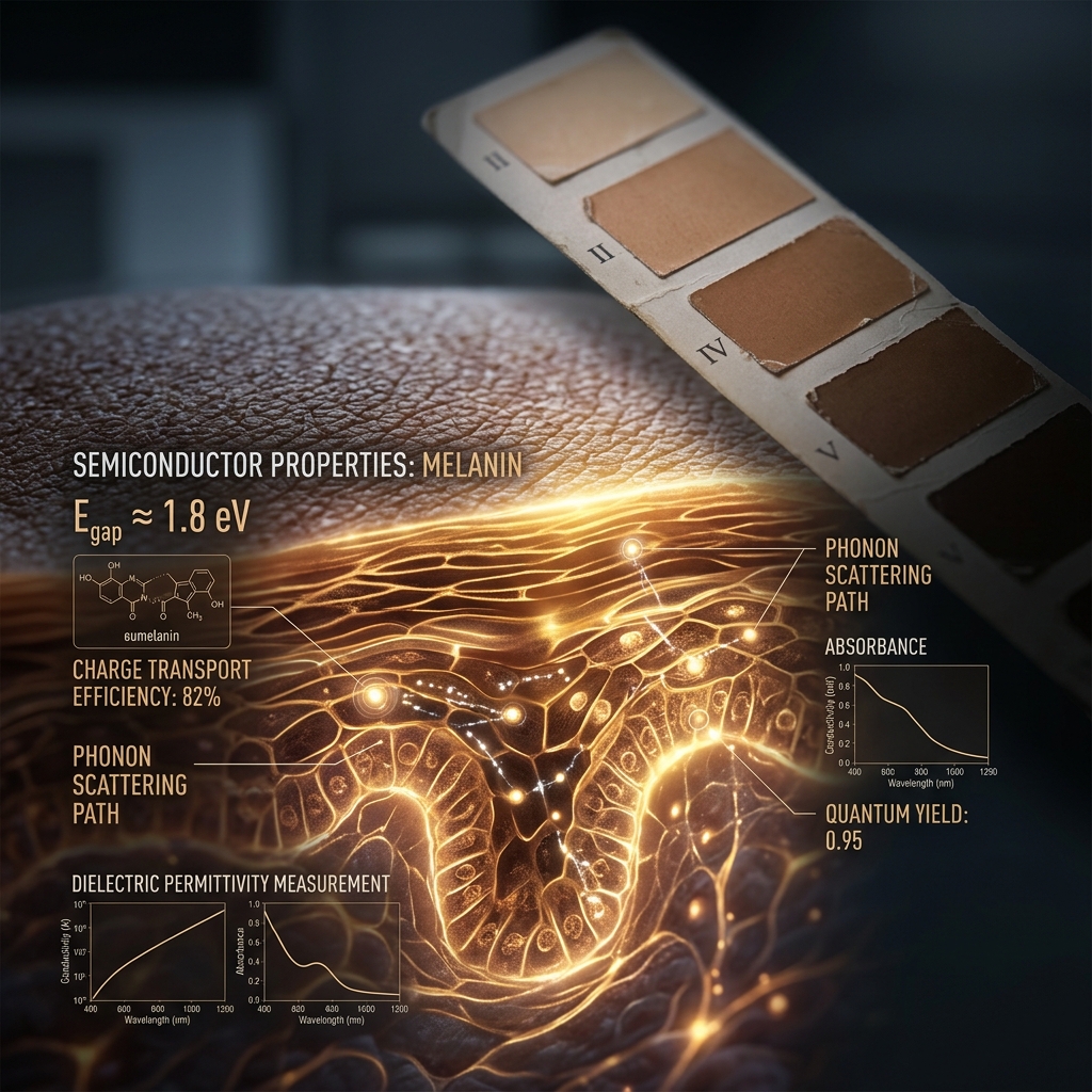
Melanin is not merely an inert pigment that imparts color; it is a dynamic, broadband radiation absorber, an amorphous organic semiconductor with a ~1.85 eV bandgap, and a hydration-dependent proton conductor. To reduce its vast biochemical and biophysical diversity to six discrete, subjective categories ignores decades of materials science and biophysics. As clinical research pushes toward personalized photomedicine, the limitations of phenotypic classification have become an active liability. A modern, melanin-based biophysical assessment framework must supersede visual typing, allowing us to interact with the integumentary system based on its true optical, chemical, and electronic properties.
The Subjectivity Trap and Optical Discordance
The core structural failure of the FST scale lies in its reliance on visual phenotyping and patient recall. Extensive clinical studies have demonstrated significant discordance between a patient's assigned Fitzpatrick type and their skin's actual optical properties, particularly in heavily pigmented populations (FST IV–VI). Dermatological studies have repeatedly shown that self-reported UV responses frequently contradict the objective melanin content of the epidermis. Visual classification conflates constitutive pigmentation (baseline genetic melanin levels) with facultative pigmentation (the UV-induced tanning response), while entirely missing the underlying physics of photon scattering.
When light or laser energy interacts with human skin, it does not interact with a "Type IV" label; it interacts with a dense, structured matrix of chromophores. The photon absorption, scattering, and subsequent thermal or electrical energy transduction are governed entirely by the physical concentration, depth, and spatial distribution of melanosomes. By clinging to a non-quantitative scale, researchers and clinicians introduce massive variability into dermatological treatments and light-based therapies. The well-documented failure of pulse oximeters to accurately read blood oxygen saturation in highly pigmented patients during the COVID-19 pandemic is a direct consequence of treating melanin as a qualitative visual trait rather than a highly specific optical filter.
Quantifying Absorption Through Diffuse Reflectance Spectroscopy
Moving beyond the FST requires measuring melanin exactly as it functions: as an optical and electronic transducer. Diffuse reflectance spectroscopy (DRS) provides a non-invasive, objective methodology to quantify the precise amount of light absorbed by epidermal melanin. By shining broad-spectrum visible and near-infrared light onto the skin and measuring the backscattered photons, DRS generates a mathematically precise absorption curve.
From these measurements, researchers calculate the Melanin Index (MI), a continuous numerical value derived from the slope of the absorption spectrum in the red and near-infrared wavelengths (typically between 650 nm and 700 nm, where hemoglobin absorption is minimal). Unlike the rigid six-point FST scale, the Melanin Index operates on a high-resolution continuum. Spectrophotometric tissue measurements have been shown to predict minimal erythema dose (MED)—the threshold for UV damage—far more accurately than self-reported surveys. By moving to MI, clinicians capture the exact optical cross-section of the tissue, ensuring that applied photomedical devices are calibrated to the patient's actual biophysics, not their perceived category.
The Critical Metric: Eumelanin to Pheomelanin Ratio
Total melanin content is only half of the biophysical equation; the chemical composition of that melanin is equally critical. Human pigmentation is determined by a varying ratio of two distinct polymers: eumelanin (brown-black) and pheomelanin (yellow-red). These two molecules behave drastically differently under photon excitation and possess different quantum mechanical profiles.
Eumelanin is a highly efficient photoprotectant. Composed of cross-linked DHI (5,6-dihydroxyindole) and DHICA (5,6-dihydroxyindole-2-carboxylic acid) oligomers, it dissipates over 99.9% of absorbed UV radiation as harmless heat through ultra-rapid internal conversion. It acts as a biological sink for free radicals and contains a stable free radical population that is readily detectable via electron paramagnetic resonance (EPR) spectroscopy.
Pheomelanin, synthesized when sulfur is incorporated into the biochemical pathway, is composed of benzothiazine derivatives. Instead of merely absorbing and safely dissipating UV radiation, pheomelanin can act as a photosensitizer. Upon UV excitation, it frequently undergoes excited-state reactions that generate reactive oxygen species (ROS), actively contributing to oxidative stress and cellular damage.
Foundational work by Shosuke Ito at Fujita Health University, utilizing high-performance liquid chromatography (HPLC) to measure degradation products—specifically pyrrole-2,3,5-tricarboxylic acid (PTCA) for eumelanin and thiazole-2,4,5-tricarboxylic acid (TTCA) for pheomelanin—demonstrated that the eumelanin-to-pheomelanin (E/P) ratio is the true determinant of a tissue's photobiological reactivity. A biophysical skin classification system must account for this ratio. Skin with high total melanin but a high pheomelanin fraction behaves entirely differently under laser excitation or UV exposure than skin with equivalent total melanin composed purely of eumelanin.
Bioelectricity and the Future of Personalized Photomedicine
The transition to biophysical skin profiling has profound implications beyond UV damage prediction, opening new frontiers in bioelectricity and quantum biology. Melanin is a unique biomaterial capable of both electron and proton conduction. Research by Paul Meredith and Tadeusz Sarna has elucidated how eumelanin acts as an amorphous semiconductor. Furthermore, its conductivity is highly dependent on hydration; water acts as a dielectric medium that modulates the mobility of protons within the melanin matrix, altering its electrochemical properties.
When we quantify skin using biophysical parameters (Melanin Index, E/P ratio, and hydration state), we can begin to predict photo-bioelectric responses. The membrane potential ($V_{mem}$) of cells, which Michael Levin's lab at Tufts University has established as crucial for directing cellular behavior, wound healing, and morphogenesis, is inevitably influenced by the local ionic and protonic environment. Because melanin serves as an endogenous proton conductor and free-radical buffer, its specific spatial and chemical distribution logically alters the electrical microenvironment of the skin.
Personalized photomedicine is currently evolving to rely on optical devices that map these exact parameters before initiating treatment. By discarding the Fitzpatrick scale in favor of continuous spectrophotometric and biochemical data, clinical science can optimize laser fluences, customize targeted photodynamic therapies, and safely explore emerging therapies utilizing exogenous electromagnetic frequencies. The future of dermatological science requires treating melanin not as a static marker of identity, but as the sophisticated, quantifiable bio-electronic interface that it truly is.
Key Takeaways
- The Fitzpatrick Skin Type (FST) scale is inherently subjective, conflates constitutive and facultative pigmentation, and frequently fails to accurately represent the optical properties of diverse skin types.
- Diffuse reflectance spectroscopy (DRS) and the continuous Melanin Index (MI) offer objective, quantitative measurements of how skin absorbs and scatters light, removing human bias from clinical assessment.
- The ratio of photoprotective eumelanin to photosensitizing pheomelanin is critical for understanding tissue reactivity; identical volumes of total melanin can yield drastically different biochemical responses to light based on this ratio.
- Eumelanin operates biologically as an amorphous semiconductor and hydration-dependent proton conductor, possessing biophysical properties that heavily influence the local cellular microenvironment.
- Transitioning to biophysical melanin assessment is vital for improving the accuracy of optical health sensors (such as pulse oximeters) and advancing the safety and efficacy of personalized photomedicine and bioelectric therapies.
References
Eilers S, Bach DQ, Gaber R, et al. "Accuracy of self-report in assessing Fitzpatrick skin phototypes I through VI." JAMA Dermatology 149(11), 1289-1294 (2013). DOI: 10.1001/jamadermatol.2013.6101.
Fitzpatrick TB. "The validity and practicality of sun-reactive skin types I through VI." Archives of Dermatology 124(6), 869-871 (1988). DOI: 10.1001/archderm.124.6.869.
Ito S, Wakamatsu K. "Quantitative analysis of eumelanin and pheomelanin in humans, mice, and other animals: a comparative review." Pigment Cell Research 16(5), 523-531 (2003). DOI: 10.1034/j.1600-0749.2003.00072.x.
Levin M. "Molecular bioelectricity: how endogenous voltage potentials control cell behavior and instruct pattern regulation in vivo." Molecular Biology of the Cell 25(24), 3835-3850 (2014). DOI: 10.1091/mbc.E13-12-0708.
Meredith P, Sarna T. "The physical and chemical properties of eumelanin." Pigment Cell Research 19(6), 572-594 (2006). DOI: 10.1111/j.1600-0749.2006.00345.x.
Stamatas GN, Zmudzka AZ, Kollias N, Beer JZ. "Non-invasive measurements of skin pigmentation in situ." Pigment Cell Research 17(6), 618-626 (2004). DOI: 10.1111/j.1600-0749.2004.00193.x.