1. Introduction to Melanin Families: Beyond Simplistic Distinctions
Eumelanin and pheomelanin are not single, chemically uniform molecules. They are heterogeneous pigment systems formed from partially oxidized, variably cross-linked building blocks and organized within melanosomes alongside proteins, lipids, ions, and water. Their measured behavior therefore depends not only on chemical composition, but also on particle size, internal packing, hydration, oxidation history, pH, metal content, and the cellular environment in which the pigment is produced.
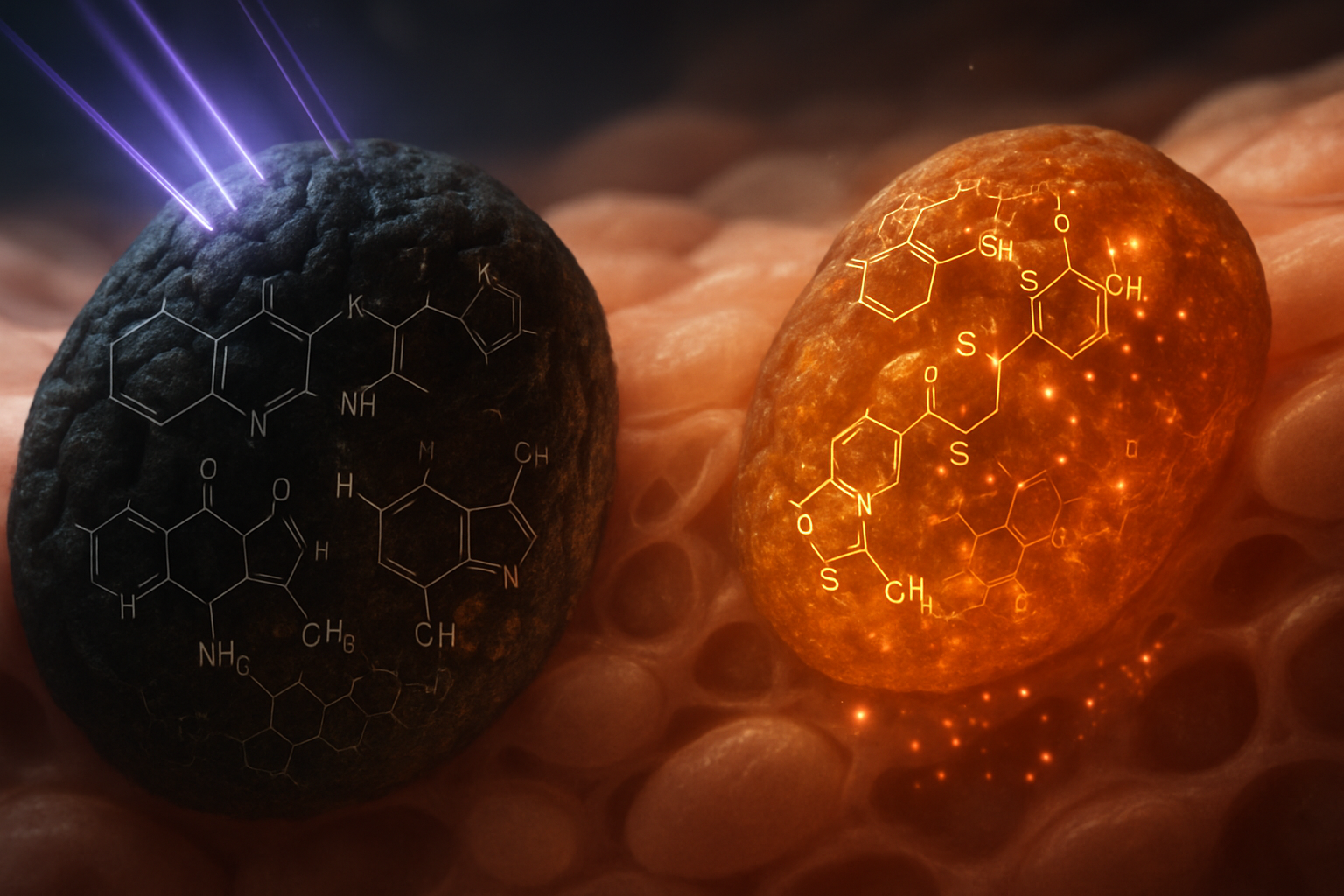
Eumelanin is usually described as a dark brown-to-black pigment composed primarily of indole-derived units related to 5,6-dihydroxyindole (DHI) and 5,6-dihydroxyindole-2-carboxylic acid (DHICA). Pheomelanin is a sulfur-containing family of pigments formed when cysteine or glutathione reacts with the common precursor dopaquinone. Its principal structural motifs are associated with benzothiazine and benzothiazole chemistry. In biological samples, these categories are not always chemically pure: mixed melanogenesis can produce particles containing both indole and sulfur-containing components, and the relative abundance of each component can vary between tissues, developmental stages, and individuals.
The distinction is therefore functional as well as structural. Eumelanin generally exhibits strong, broadband absorption extending from the ultraviolet into the visible region, efficient conversion of absorbed photon energy into vibrational heat, and very weak fluorescence. Pheomelanin also absorbs ultraviolet radiation, but its sulfur-containing chromophores can introduce different electronic states and redox pathways. Under particular conditions—especially in the presence of oxygen, transition metals, or continued oxidative chemistry—these pathways may increase the likelihood of reactive oxygen species formation and persistent photochemical reactions.
A useful comparison is not simply “dark pigment versus light pigment.” It is a comparison between two distributed molecular systems that differ in:
- the identity and oxidation state of their aromatic building blocks;
- the presence or absence of sulfur-containing units;
- the balance of DHI and DHICA-like components;
- the degree of aggregation and π-electron coupling;
- the density of radical and quinone sites;
- the ability to bind metals and protons;
- the response to hydration and pH; and
- the relative rates of energy dissipation, electron transfer, and reactive oxygen chemistry.
These variables are interconnected. For example, a carboxyl group in DHICA can increase hydrophilicity and metal-binding capacity while also modifying intermolecular packing. Hydration can alter electronic coupling between pigment units and can change electrical conductivity by several orders of magnitude in synthetic eumelanin films, depending on the measurement method and humidity range. Oxidation can create quinone sites that accept electrons, whereas reduction can regenerate hydroquinone-like sites capable of donating them. The pigment is consequently better understood as a redox-active, disordered organic material than as an inert colorant.
The central biophysical question is how absorbed energy and charge are handled. In a protective configuration, excitation is rapidly dissipated as heat and radicals are quenched or stabilized. In a chemically permissive configuration, the same broad electronic structure can support electron transfer, oxygen reduction, lipid oxidation, or delayed photochemical reactions. The balance is influenced by DHI/DHICA composition, sulfur content, hydration, pH, particle morphology, and metal occupancy.
2. The Chemistry of Melanin Synthesis: A Diversion in Pathways
The Critical Dopaquinone Intersection
Melanin formation begins with the hydroxylation of L-tyrosine to L-3,4-dihydroxyphenylalanine (L-DOPA) and the subsequent oxidation of L-DOPA to dopaquinone. In melanocytes, these reactions are initiated and accelerated by tyrosinase, a copper-containing oxidase. Dopaquinone is a highly electrophilic intermediate and represents the principal chemical branch point between eumelanin- and pheomelanin-dominant synthesis.
When cysteine or another available sulfur nucleophile is abundant, it attacks dopaquinone rapidly. The initial products include cysteinyldopa isomers, particularly 5-S-cysteinyldopa and 2-S-cysteinyldopa. These intermediates undergo oxidation, cyclization, decarboxylation, and rearrangement to produce benzothiazine-related structures. Further oxidation and chemical transformation can generate benzothiazole-containing units. The precise distribution depends on pH, oxygen availability, the cysteine:dopaquinone ratio, enzyme activity, and the duration of oxidation.
When sulfur nucleophiles are relatively depleted, dopaquinone undergoes intramolecular cyclization to form dopachrome. Dopachrome is itself a branching intermediate. It can decarboxylate to form DHI or undergo rearrangement toward DHICA. Two enzymes are particularly important in this context:
- Tyrosinase-related protein 2, also called dopachrome tautomerase, favors the conversion of dopachrome toward DHICA.
- Tyrosinase-related protein 1 participates in downstream oxidation reactions, although its precise contribution depends on substrate, pH, and cellular context.
This means that eumelanin composition is not determined only by whether melanogenesis is “on.” It is also shaped by the relative rates of dopachrome decarboxylation, tautomerization, oxidation, and oligomerization.
The sulfur branch is similarly conditional. Cysteine concentrations inside melanosomes can be transiently high during active melanogenesis, while glutathione may act as an additional sulfur source. As dopaquinone production increases, the local rate of quinone formation can exceed the rate at which sulfur nucleophiles are supplied. A cell may therefore produce a mixture of sulfur-containing and indole-derived pigments even without a categorical genetic switch between pheomelanogenesis and eumelanogenesis.
The intracellular compartment matters as much as the chemical reaction. Melanosomes provide a partially isolated reaction volume with controlled pH, protein scaffolding, membrane-associated transport, and restricted diffusion. Early-stage melanosomes are more acidic and less heavily pigmented, whereas more mature melanosomes become increasingly electron-dense and optically absorbing. Changes in organelle pH affect tyrosinase activity, cysteine availability, quinone reactivity, and the stability of intermediates. Consequently, two pigment samples with similar bulk sulfur content can display different optical or redox behavior if they were assembled under different conditions.
The Distinctive Roles of DHI and DHICA in Eumelanin
DHI and DHICA share an indole-based aromatic framework but differ substantially in charge distribution and intermolecular behavior. DHI contains two phenolic hydroxyl groups and an indole nitrogen, whereas DHICA additionally contains a carboxylic acid at the 2-position. At physiological and mildly alkaline pH, the DHICA carboxyl group can be deprotonated, giving the unit a negative charge. This increases hydration and creates potential coordination sites for metal ions such as iron, copper, calcium, and zinc.
DHI-rich material generally forms more compact, strongly absorbing aggregates. DHI can readily undergo oxidation to quinone and quinone-methide-like states, followed by coupling reactions that produce oligomeric and polymeric structures. DHICA-containing material is often more polar and can adopt different aggregation geometries. The negatively charged carboxylate groups introduce electrostatic repulsion between units and can reduce the tendency of aromatic surfaces to pack as tightly as they would in a neutral DHI-rich assembly. The result is not necessarily a simple “more ordered” or “less ordered” material; rather, DHICA changes the balance between π–π interactions, hydrogen bonding, ion pairing, hydration, and metal coordination.
A worked conceptual example illustrates this effect. Consider two hypothetical pigment particles with the same total number of indole units:
- Particle A contains a high proportion of DHI-like units. Its neutral aromatic surfaces can approach closely, increasing π-electron coupling and creating dense, strongly absorbing domains.
- Particle B contains a larger fraction of DHICA-like units. Its surface includes more carboxylates, which bind water and may coordinate metal ions. The increased hydration shell can separate neighboring chromophores by fractions of a nanometer, modifying exciton coupling and charge-hopping pathways.
The particles may have similar visible color but differ in conductivity, metal-binding behavior, swelling, and response to pH. In a low-humidity state, their electrical properties may converge toward those of a poorly conducting amorphous solid. As humidity rises, Particle B may show a stronger environmental response because its ionizable groups support proton and ion mobility.
The DHI/DHICA ratio is therefore a structural variable with multiple consequences. It can affect:
- Optical absorption: changes in conjugation, aggregation, and the distribution of electronic transition energies.
- Acid-base behavior: carboxylates add titratable sites and alter the effective isoelectric behavior of the material.
- Metal binding: carboxylate and phenolate groups can coordinate cations.
- Hydration: charged DHICA-rich surfaces generally bind more water.
- Mechanical properties: hydrogen bonding, ionic interactions, and hydration influence particle rigidity and swelling.
- Charge transport: localized states and hopping distances change with packing and water content.
The DHI/DHICA ratio also varies across species and tissues. In addition, chemical degradation during extraction can alter the apparent ratio, making comparisons between synthetic, isolated, and native pigment difficult unless the preparation and analytical method are carefully controlled.
Utilization of Benzothiazine and Benzothiazole in Pheomelanin
Pheomelanin formation begins with sulfur addition to dopaquinone. The resulting cysteinyldopa intermediates retain sulfur in a chemically active position that can participate in ring closure and oxidation. Benzothiazine structures are generally regarded as relatively early or less extensively oxidized pheomelanin-related motifs, while benzothiazole structures represent more oxidized and rearranged products. Native pheomelanin is likely to contain a distribution of related structures rather than a regular repeating polymer.
Sulfur changes the electronic landscape in several ways. Sulfur atoms are larger and more polarizable than oxygen, and their lone-pair electrons can participate in charge-transfer interactions and redox chemistry. Sulfur-containing heterocycles also introduce additional sites at which oxidation can occur. Depending on the local environment, sulfur may exist in reduced thioether-like forms, oxidized states, or structures integrated into aromatic heterocycles. These states do not all have the same electron-donating ability or photochemical behavior.
Pheomelanin's sulfur content is frequently discussed in relation to its lower photoprotective efficiency compared with eumelanin, but the explanation is not simply that sulfur “absorbs more light.” The relevant issue is what happens after absorption. A chromophore that rapidly relaxes to the ground state through internal conversion primarily produces heat. A chromophore that supports longer-lived charge-separated states or efficient intersystem crossing can transfer electrons or energy to molecular oxygen. Pheomelanin may provide more opportunities for the latter processes, especially when partially oxidized sulfur and quinone groups coexist.
The chemical environment remains decisive. A dry pheomelanin preparation, a hydrated pheomelanin particle inside a melanosome, and pheomelanin associated with membrane lipids can have different redox potentials and different access to oxygen. Lipid association may place reactive intermediates close to unsaturated fatty acids, while protein association may restrict diffusion or stabilize specific radical states. Thus, the presence of pheomelanin is a risk factor for particular chemistries, not a guarantee that every pheomelanin-containing system will behave as a strong pro-oxidant.
Shared Redox Characteristics
Both pigment families contain mixtures of reduced and oxidized sites. Their redox chemistry can be represented conceptually by interconversion among hydroquinone-like, semiquinone, and quinone states:
[ \text{hydroquinone} \rightleftharpoons \text{semiquinone radical} \rightleftharpoons \text{quinone} ]
This is not a single equilibrium for the whole pigment. It is a distribution of local states with different redox potentials. A pigment particle may simultaneously contain electron-rich phenolic groups, semiquinone radicals, oxidized quinones, carboxylates, bound metal ions, and trapped charges.
The semiquinone state is particularly important because it explains why melanins can display persistent electron paramagnetic resonance signals. These radicals are often relatively stable because the unpaired electron is delocalized over an aromatic framework and because the disordered matrix limits the bimolecular reactions that would otherwise rapidly eliminate radicals. Persistent, however, does not mean chemically inert. A semiquinone can donate an electron to an acceptor such as oxygen or receive an electron from a reducing agent such as glutathione, depending on the local potential.
Melanin can therefore behave as:
- an electron donor that reduces metal ions or reactive intermediates;
- an electron acceptor that oxidizes cellular reductants;
- a radical sink that stabilizes unpaired electrons;
- a metal ligand that changes metal-centered redox chemistry; or
- a redox mediator that transfers charge between otherwise separated molecules.
The outcome depends on pH, oxygen tension, hydration, metal concentration, and the ratio of reduced to oxidized sites. At low oxygen availability, melanin may mainly buffer radicals. In oxygen-rich conditions, the same reduced sites may facilitate oxygen reduction and generate superoxide or hydrogen peroxide. A transition metal can further amplify this chemistry; for example, iron cycling between Fe²⁺ and Fe³⁺ can couple pigment electron transfer to peroxide chemistry and, under suitable conditions, hydroxyl-radical formation.
3. Melanin's Physical Assembly: From Molecular Structures to Material Properties
The Eumelanin Electronic Network
Modern descriptions of eumelanin increasingly treat it as a disordered electronic ensemble rather than a conventional, regularly repeating polymer. Small indole-derived oligomers can stack or associate into nanoscale domains, while larger aggregates contain a distribution of structural defects, oxidation states, hydration pockets, and ionic sites. The resulting material has no single band gap or sharply defined set of molecular orbitals. Instead, it contains a broad distribution of localized electronic states.
This picture helps explain several characteristic observations:
- Eumelanin absorbs broadly rather than displaying only a few narrow spectral peaks.
- Its conductivity is usually low in dry conditions but can increase dramatically with humidity.
- Its electron paramagnetic resonance spectrum often contains a relatively persistent signal.
- Its optical properties vary with oxidation state, pH, particle size, and preparation history.
- Different laboratories may obtain different conductivity or absorption values from nominally similar samples.
In an ordered semiconductor, charge carriers may move through extended bands. In eumelanin, charge is more often described by localized-state transport, including thermally assisted hopping between chemically or spatially separated sites. The distance between sites, their energetic disorder, and the availability of protons or counterions all affect the measured current. Protonic conduction can become particularly important under humid conditions, where water forms transient pathways through hydrophilic regions.
A simple worked comparison can clarify the environmental dependence. A dry synthetic eumelanin film measured at low relative humidity may show current close to the instrument's detection limit at modest applied voltage. If the same film is equilibrated at high relative humidity, absorbed water can plasticize the matrix, increase ion mobility, alter protonation states, and create additional hopping pathways. The current may rise by orders of magnitude even though the chemical identity of the pigment has not changed. The material has not become a crystalline conductor; rather, water has changed the availability and connectivity of localized charge-transfer pathways.
At the nanoscale, eumelanin is often described as consisting of stacked or associated oligomeric units with dimensions on the order of a few nanometers embedded within larger particles. Exact structural dimensions depend strongly on the technique used—electron microscopy, scanning probe microscopy, scattering, spectroscopy, or computational modeling—and should not be interpreted as a single universal architecture. What is robust is the presence of hierarchical organization: molecular units form small electronic domains, which are assembled into larger pigment granules and ultimately into melanosomes that may be approximately 0.5–1 micrometer in size.
The Nature of Pheomelanin Aggregates
Pheomelanin aggregates are also heterogeneous, but sulfur-containing motifs introduce additional variation in polarizability, oxidation chemistry, and intermolecular interactions. Benzothiazine and benzothiazole-related units may not stack or pack in the same way as DHI- and DHICA-rich units. Sulfur can participate in dispersion interactions with aromatic surfaces, while the polar and redox-active nature of the heterocycles can increase sensitivity to oxygen, pH, and surrounding biomolecules.
Compared with eumelanin, pheomelanin often displays a more complex relationship between composition and optical response. The apparent color of a pheomelanin-containing sample is determined by the overlap of many broad transitions, by particle scattering, and by the proportions of different sulfur-containing structures. Oxidation can shift absorption and change the relative contribution of benzothiazine- and benzothiazole-like motifs. Consequently, two reddish-yellow or brown pigment samples may differ substantially in radical persistence and photochemical activity.
Pheomelanin aggregates can also be influenced by associated lipids and proteins. In biological melanosomes, pigment is not a free powder; it is deposited on a proteinaceous matrix and enclosed by a membrane. Sulfur-containing pigment may therefore be positioned near membrane lipids or redox-active proteins. The local arrangement determines whether a photogenerated radical is rapidly trapped within the pigment, transferred to oxygen, or passed to a neighboring lipid or protein.
The aggregate's physical size is also important. Smaller particles have a larger surface-to-volume ratio, making a greater fraction of their reactive groups accessible to water, oxygen, and metal ions. Larger, densely packed particles may shield internal sites but can retain long-lived trapped charges. Surface chemistry can therefore dominate biological interactions even when the bulk elemental composition is similar.
Intrinsic Disorder as Functionality
Disorder in melanin is not merely a manufacturing defect. It is a source of broad spectral absorption, multiple redox potentials, and adaptive responses to the environment. In a structurally heterogeneous particle, photons of different energies can be absorbed by different subpopulations of chromophores. Excited states can then migrate over short distances before being trapped or converted into vibrational energy.
The same disorder creates a landscape of localized charge sites. Charge may move through a sequence of thermally activated hops:
- an electron or hole is generated at an oxidized or excited chromophore;
- it migrates to a nearby site with a compatible energy;
- local molecular motion or hydration assists the transfer;
- the charge becomes trapped at a lower-energy site or continues to another domain.
The time scale and distance of each hop are variable. There is no requirement for a charge to travel through a perfectly periodic lattice. This is one reason melanin can show both insulating and semiconducting-like behavior depending on frequency, temperature, humidity, and applied voltage.
Water is especially influential. It changes hydrogen bonding, protonation, dielectric screening, and molecular mobility. A pigment's response to an alternating electric field can consequently differ from its response to a direct current. At low frequencies, ions and protons may move over relatively long distances; at higher frequencies, only local polarization and short-range charge displacement may respond. These distinctions are important when interpreting claims about “conductivity” or “semiconductivity,” because different measurement regimes may probe different physical mechanisms.
4. Photophysical Properties: Light Interaction and Consequences
Eumelanin's UV-Visible Roles
Eumelanin absorbs strongly across a broad spectral range, with particularly important protection in the ultraviolet. Its absorption does not arise from one isolated chromophore but from overlapping transitions associated with differently oxidized indole units, oligomeric domains, and aggregate-induced electronic states. The absorption coefficient generally decreases gradually toward longer wavelengths, producing the characteristic dark, broadband appearance.
A key protective property is the high efficiency of nonradiative decay. Fluorescence quantum yields for eumelanin are commonly very low—often reported in the range of approximately (10^{-4}) to (10^{-3}), depending on sample preparation and measurement conditions—indicating that only a small fraction of absorbed photon energy is released as fluorescence. Instead, the energy is converted into vibrational motion of the pigment and surrounding matrix.
If a photon in the ultraviolet carries approximately 4–5 electronvolts of energy, eumelanin can dissipate that energy through a cascade of internal conversion and vibrational relaxation events. At the scale of a melanosome, the final result is a small increase in local heat rather than the emission of a high-energy photon or the creation of a long-lived excited state. The effectiveness of this process is one reason melanin can protect nucleic acids and proteins from direct ultraviolet excitation.
Protection also depends on spatial organization. Melanosomes absorb and scatter light before it reaches the nucleus. Their size, shape, density, and distribution within keratinocytes determine optical path length and shielding. Larger, more elongated melanosomes tend to resist rapid degradation and can provide prolonged optical protection, whereas smaller or less densely packed melanosomes may offer less effective attenuation even if their pigment chemistry is similar.
Nonradiative Relaxation's Dominance
Excited-state relaxation in eumelanin is unusually rapid. Ultrafast spectroscopy commonly identifies components in the femtosecond-to-picosecond regime, although slower processes can persist from nanoseconds to much longer times as charges become trapped in the disordered matrix. The initial rapid component reflects internal conversion and vibrational redistribution; the slower components may involve localized polarons, radical pairs, structural relaxation, or diffusion of energy and charge among pigment domains.
The existence of a fast component does not mean every excitation follows exactly one pathway. Eumelanin contains many chromophores and environments, so the observed signal is usually multiexponential or broadly distributed. Some sites may relax in less than a picosecond, while others support longer-lived trapped states. The important functional point is that the dominant early-time pathway is strongly nonradiative.
This can be expressed using a qualitative rate relationship:
[ \Phi_{\mathrm{heat}} \approx \frac{k_{\mathrm{IC}}}{k_{\mathrm{IC}}+k_{\mathrm{fl}}+k_{\mathrm{ISC}}+k_{\mathrm{ET}}} ]
where (k_{\mathrm{IC}}) is the internal-conversion rate, (k_{\mathrm{fl}}) is the fluorescence rate, (k_{\mathrm{ISC}}) is the intersystem-crossing rate, and (k_{\mathrm{ET}}) represents electron-transfer pathways. In eumelanin, the effective internal-conversion contribution is large, while fluorescence is small. The balance can nevertheless shift with oxidation state, hydration, metal binding, and aggregation.
Hydration can either improve or complicate photoprotection. It may facilitate vibrational relaxation and stabilize charge-separated states, but it can also increase molecular mobility and permit electron transfer to dissolved oxygen or nearby biomolecules. Thus, the same water content that enhances a pigment's electrical response may alter its photochemical response.
Pheomelanin's Excited-State Pathways
Pheomelanin also absorbs ultraviolet radiation and can dissipate some excitation as heat, but its sulfur-containing units provide alternative channels for excited-state evolution. Sulfur can enhance spin-orbit coupling relative to oxygen-containing analogues, potentially increasing access to triplet-like states or other excited configurations. The exact importance of this effect varies with the molecular structure and oxidation state of the pigment, but it provides a plausible route by which excitation can interact more efficiently with oxygen.
Two broad photochemical processes are relevant:
- Electron transfer: an excited pigment unit may donate an electron to oxygen or accept one from a neighboring donor, producing superoxide, semiquinone, or other radical species.
- Energy transfer: an excited state may transfer energy to oxygen, potentially generating electronically excited oxygen species such as singlet oxygen.
The resulting chemistry is highly context-dependent. Oxygen concentration, antioxidant availability, pigment hydration, and the proximity of lipids all influence the outcome. A pheomelanin particle surrounded by glutathione may be kept in a more reduced state, whereas one near oxidized lipids and catalytic metal ions may support sustained radical chemistry.
Pheomelanin can therefore be photochemically active even after the initiating ultraviolet photon has disappeared. Long-lived intermediates may continue to react on microsecond, millisecond, or still longer time scales, particularly if the pigment contains trapped charges or quinone-like sites. This does not imply that all pheomelanin is intrinsically destructive. Rather, it means that its excited-state pathways may be less exclusively committed to ultrafast heat production than those of a strongly protective eumelanin-rich system.
Investigating Photophysical Outcomes
The difference between protective and damaging photophysics is best examined using several complementary measurements rather than color alone. Useful approaches include:
- Transient absorption spectroscopy, which follows excited states and charge-separated intermediates from femtoseconds to milliseconds.
- Time-resolved fluorescence, which measures the small radiative component and its lifetime.
- Electron paramagnetic resonance, which detects persistent and light-induced radicals.
- Singlet-oxygen and superoxide assays, which probe reactive oxygen production but require careful controls for dye sensitization and assay interference.
- Oxygen-consumption measurements, which determine whether irradiation accelerates redox reactions.
- DNA photoproduct analysis, which reveals whether chemically damaging products appear during or after irradiation.
- Lipid-peroxidation assays, which test whether pigment-associated radicals propagate into membranes.
A particularly important observation in pheomelanin biology is delayed photochemistry. Studies of red-hair/fair-skin models and isolated pigment systems have shown that oxidative reactions initiated by ultraviolet exposure can continue after the exposure period. In the chemiexcitation mechanism, redox reactions generate high-energy carbonyl species whose energy can be transferred to melanin-related molecules, producing electronically excited states without direct absorption of a new photon. These excited states can then promote DNA photoproduct formation in the dark.
The practical implication is that the end of ultraviolet exposure does not necessarily mark the end of photochemical risk. The magnitude and duration of this effect depend on pigment composition, cellular antioxidants, DNA proximity, temperature, oxygen, and the efficiency of repair pathways. Eumelanin-rich systems generally show stronger photoprotective behavior, but eumelanin is not chemically invulnerable; heavily oxidized pigment, metal-loaded pigment, or pigment exposed to intense irradiation can also participate in pro-oxidant reactions.
5. Redox Dynamics and Pigment Function: Antioxidant vs. Pro-Oxidant Performance
The Redox Flexibility of Melanin
Melanin's redox flexibility derives from the coexistence of phenols, catechols, quinones, semiquinone radicals, indole nitrogens, carboxylates, sulfur-containing groups, and bound metal ions. Instead of having one fixed oxidation-reduction potential, a pigment particle contains a broad ensemble of sites with different electron affinities.
This distributed chemistry allows melanin to interact with reactive species through several mechanisms:
- direct electron transfer;
- hydrogen-atom transfer;
- radical addition to quinone sites;
- metal-ion chelation;
- proton-coupled electron transfer; and
- physical confinement of radicals within the pigment matrix.
A pigment can consume a radical without fully eliminating oxidative capacity. For example, an antioxidant molecule may reduce a quinone to a semiquinone or hydroquinone state, while the pigment later transfers an electron to oxygen and regenerates a more oxidized form. This redox cycling can be protective if it terminates reactive intermediates, but it can become pro-oxidant if oxygen reduction is efficient and antioxidant replenishment is limited.
The biological outcome is consequently determined by flux, not just by the presence of a reactive group. A pigment that has many radical sites may act as a stable radical reservoir with little net damage, whereas a pigment with fewer but more accessible sites may catalyze repeated electron-transfer cycles.
Eumelanin's Role in Redox Balancing
Eumelanin can scavenge several reactive species, including carbon-centered radicals and some oxygen-derived radicals. Its aromatic and phenolic structure provides multiple routes for delocalizing the resulting unpaired electron. In addition, its metal-binding sites can sequester iron and copper, reducing the availability of free metal ions for uncontrolled peroxide chemistry.
However, metal binding is not automatically protective. A tightly coordinated metal may be chemically inert, while a partially accessible metal center can facilitate electron transfer between pigment and oxygen. The difference depends on coordination geometry, oxidation state, ligand exchange, and local hydration. For example, an iron ion bound within a relatively dry, shielded pigment domain may be less reactive than an iron ion positioned at a hydrated particle surface.
pH also changes eumelanin redox behavior. Deprotonation of phenolic and carboxylic groups alters electron density and metal affinity. At higher pH, phenolate formation can make electron donation easier but can also increase the rate of oxidation to quinones. At lower pH, protonation changes the balance of hydrogen bonding and can affect the stability of semiquinone states. Melanosomal pH is therefore relevant not only to tyrosinase activity but also to the mature pigment's subsequent redox behavior.
Hydration creates another balance. A hydrated eumelanin matrix can accept and transport protons, supporting proton-coupled electron transfer. Water may also increase the mobility of oxygen and small reductants. The consequence can be faster radical neutralization or faster redox cycling, depending on which reactants are available.
Eumelanin's protection is also physical. By absorbing ultraviolet light and distributing excitation energy over many coupled sites, it lowers the probability that a single DNA molecule or membrane lipid receives the full energy of an absorbed photon. The pigment can thus combine optical shielding, ultrafast heat dissipation, radical stabilization, and metal sequestration.
Sulfur-Influenced Dynamics in Pheomelanin
Sulfur-containing pheomelanin introduces chemically accessible atoms and heterocycles that can participate in oxidation and electron transfer. Sulfur may be more readily oxidized than a fully aromatic carbon framework, and oxidation can generate sulfoxide-, sulfone-, or sulfur-centered radical chemistry depending on the precise structure. The resulting products can alter polarity, charge distribution, and oxygen reactivity.
This is one reason pheomelanin has been associated with increased oxidative stress in several experimental systems. Under ultraviolet illumination, pheomelanin can promote formation of reactive oxygen species and may contribute to oxidative damage in nearby DNA and lipids. In red-hair/fair-skin biological models, pheomelanin-associated oxidative stress has also been linked to melanoma susceptibility through mechanisms that are not entirely dependent on direct ultraviolet exposure. The effect is likely influenced by both the pigment itself and the broader cellular state, including antioxidant capacity, inflammation, mitochondrial metabolism, and DNA repair.
A useful conceptual comparison is a redox cycle involving a pigment and oxygen:
[ \text{Pheo}{\mathrm{red}} + \mathrm{O_2} \rightarrow \text{Pheo}{\mathrm{ox}} + \mathrm{O_2^{\bullet -}} ]
The superoxide produced in such a schematic reaction can dismutate, spontaneously or through superoxide dismutase, to hydrogen peroxide. Hydrogen peroxide can then be removed by catalase, glutathione peroxidase, or peroxiredoxins—or can participate in metal-mediated chemistry if reactive iron or copper is available. Whether the net effect is damaging depends on the rates of each competing reaction.
Pheomelanin may also act as a sink for cellular reductants. Glutathione, ascorbate, and NAD(P)H-linked systems can reduce oxidized pigment sites, but continuous reoxidation by oxygen may consume antioxidant equivalents. This creates a possible mechanism by which pigment-associated redox cycling contributes to oxidative burden even in the absence of intense external irradiation.
Again, composition matters. Benzothiazole-rich and benzothiazine-rich fractions may differ in redox potential, photostability, and radical persistence. Pheomelanin extracted by harsh chemical procedures may also undergo oxidation or fragmentation that changes its behavior relative to native melanosomes. Claims about “pheomelanin” should therefore specify whether they refer to intact organelles, synthetic model pigments, isolated natural pigment, or chemically oxidized material.
Towards Predictive Models of Pigment Activity
Predicting pigment behavior requires connecting structural diagnostics to measurable function. A useful model would include at least the following variables:
- elemental sulfur content and sulfur speciation;
- DHI/DHICA abundance;
- quinone-to-hydroquinone balance;
- radical concentration and persistence;
- particle size and surface area;
- hydration-dependent conductivity;
- bound iron, copper, and other metals;
- local pH and ionic strength;
- oxygen accessibility; and
- proximity to DNA, proteins, and unsaturated lipids.
The same pigment can switch functional character when these parameters change. For example, a dry, metal-poor eumelanin film may mainly function as an optical absorber, while the hydrated and metal-loaded form may additionally behave as a redox-active electrochemical material. A pheomelanin-containing melanosome with abundant glutathione may show limited net oxidation, whereas depletion of glutathione can expose its capacity for oxygen reduction and delayed photochemistry.
Analytical strategies should therefore combine spectroscopy, electrochemistry, microscopy, and biochemical assays. Raman and infrared methods can report on functional groups; X-ray absorption and related methods can probe metal coordination and sulfur oxidation; electron paramagnetic resonance can quantify radical populations; impedance spectroscopy can resolve hydration- and frequency-dependent charge transport; and ultrafast optical techniques can separate early energy dissipation from longer-lived charge processes.
Predictive modeling could eventually support pigment engineering. Adjusting the DHI/DHICA ratio, controlling sulfur incorporation, removing catalytic metals, or modifying particle hydration could produce melanin-based materials optimized for ultraviolet shielding, redox buffering, biosensing, or electronic interfaces. In medicine, the same framework may help distinguish when pigment chemistry is protective from when it contributes to oxidative damage. In technology, it could guide the design of environmentally responsive coatings, biodegradable conductors, and light-absorbing biomimetic materials.
6. Synthesis of Key Insights
Eumelanin and pheomelanin are pigment collectives whose structural differences produce distinct but overlapping biophysical outcomes. Eumelanin is dominated by indole-derived chemistry involving DHI and DHICA, whereas pheomelanin incorporates sulfur through benzothiazine- and benzothiazole-related structures generated from cysteine or glutathione reactions with dopaquinone.
The difference between them is expressed across several levels:
- Chemical: sulfur incorporation changes oxidation pathways, polarizability, and available redox sites.
- Molecular: DHI and DHICA differ in charge, hydration, stacking, and metal-binding behavior.
- Supramolecular: disordered oligomeric domains assemble into pigment particles with variable density and surface chemistry.
- Photophysical: eumelanin generally favors ultrafast nonradiative energy dissipation, while pheomelanin may provide more opportunities for electron transfer, oxygen activation, and delayed chemiexcitation.
- Redox: both pigments can scavenge radicals or participate in redox cycling, with the outcome determined by pH, hydration, oxygen, metals, and antioxidant availability.
- Biological: melanosome architecture, intracellular location, pigment distribution, and DNA proximity determine whether a given chemical pathway is protective or damaging.
Eumelanin's broad absorption and efficient conversion of excitation energy into heat make it an effective photoprotective material. Its disordered structure also allows it to stabilize radicals, bind metals, and respond to changes in hydration. These same properties can support redox cycling under conditions of high oxidative pressure.
Pheomelanin's sulfur-containing chemistry gives it a different balance of excited-state and redox pathways. It may be less effective as a purely dissipative photoprotective material and more capable of supporting persistent oxidative reactions, including reactions that continue after ultraviolet exposure has ended. Yet pheomelanin behavior is not uniform or inevitably harmful. Its activity depends on structural composition, oxidation state, antioxidant buffering, metal occupancy, and the organization of the melanosome.
The major challenge is therefore to correlate molecular heterogeneity with biological function. Bulk pigment labels are insufficient unless they are accompanied by measurements of composition, hydration, oxidation state, particle morphology, radical concentration, and associated biomolecules. Resolving these relationships will be important for understanding pigmentation, melanoma susceptibility, oxidative stress, and the design of melanin-inspired materials. For the Quantum Melanin Research Foundation and related research communities, the central opportunity lies in treating melanin as a dynamic biological material—one capable of converting photons, protons, electrons, and chemical free energy in ways governed by nanoscale structure and cellular context.
Related Research
- [Melanin as a Biological Semiconductor: Bridging Quantum Physics and Cellular Biology](/articles/melanin-as-a-biological-semiconductor-bridging-quantum-physics-and-cellular-biology-1786976946135)
- Frequency-Dependent Biological Responses: Insights from Electromagnetic Stimulation of Living Systems
- The Evolutionary Significance of Melanin: Why Nature’s Most Ancient Pigment Persists Across Life
References
- Meredith, P.; Sarna, T. "The Physical and Chemical Properties of Eumelanin." Pigment Cell Research (2006). doi:10.1111/j.1600-0749.2006.00345.x
- Meredith, P.; Powell, B. J.; Riesz, J.; Nighswander-Rempel, S. P.; Pederson, M. R.; Moore, E. G. "Towards Structure–Property–Function Relationships for Eumelanin." Soft Matter (2006). doi:10.1039/b511922g
- d’Ischia, M.; Wakamatsu, K.; Napolitano, A.; Briganti, S.; García-Borrón, J. C.; Bastiaens, M.; et al. "Melanins and Melanogenesis: From Pigment Cells to Human Health and Technological Applications." Pigment Cell & Melanoma Research (2015). doi:10.1111/pcmr.12393
- Mitra, D.; Luo, X.; Morgan, A.; Wang, J.; Hoang, M. P.; Lo, J.; et al. "An Ultraviolet-Radiation-Independent Pathway to Melanoma in the Red Hair/Fair Skin Background." Nature (2012). doi:10.1038/nature11624
- Premi, S.; Wallhäuser, J.; Botti, E.; et al. "Chemiexcitation of Melanin Derivatives Induces DNA Photoproducts Long after UV Exposure." Science (2015). doi:10.1126/science.1256022