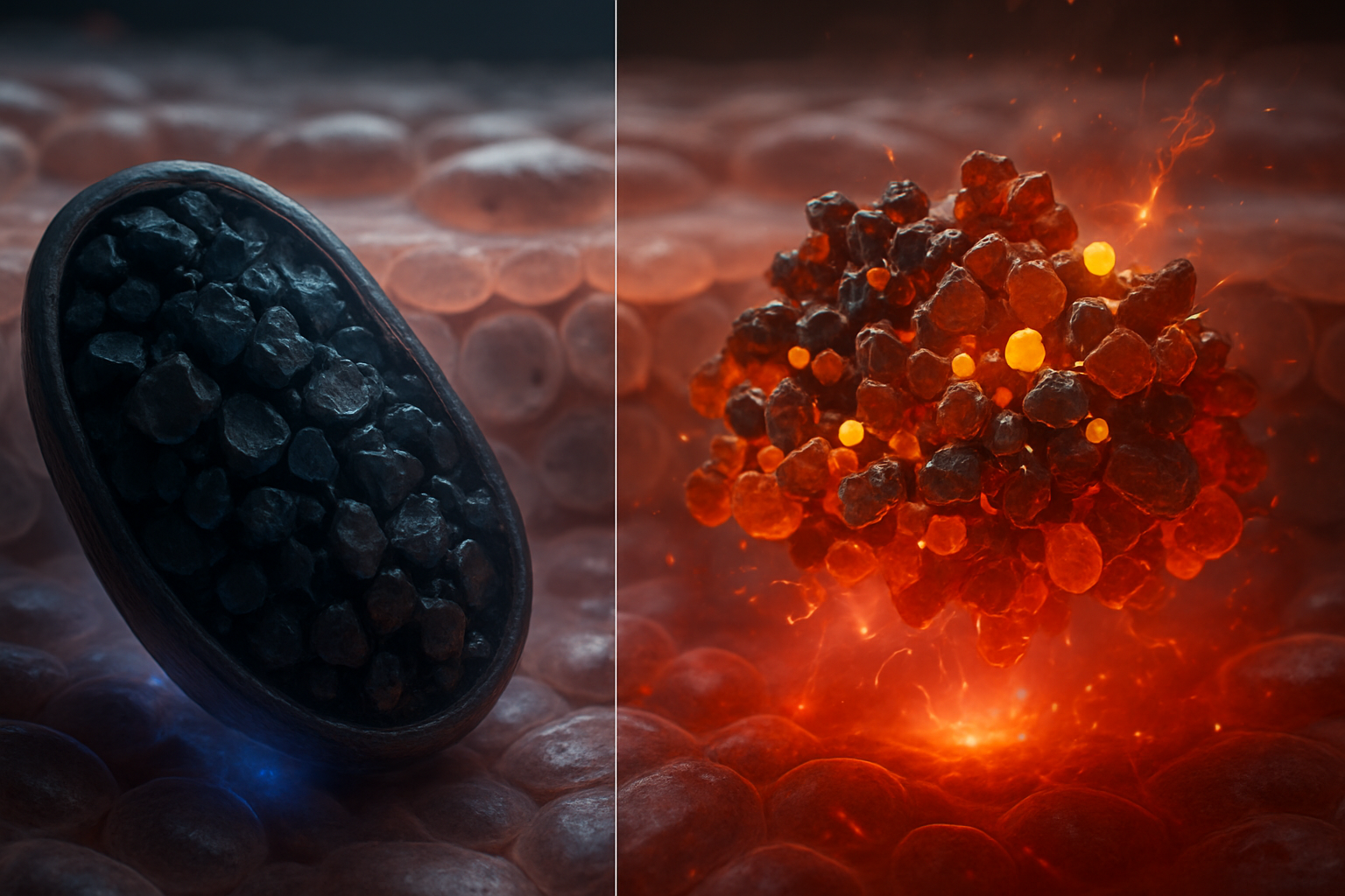
Introduction: Two Melanins, Two Unique Biophysical Profiles
Eumelanin and pheomelanin, although chemically related, are distinct pigment families with unique properties that are fundamental to many biological processes. These differences are inherently tied to their biosynthetic origins, structural composition, optical features, charge transport mechanisms, redox activities, and interactions with cellular systems. Eumelanin predominantly derives from 5,6-dihydroxyindole (DHI) and 5,6-dihydroxyindole-2-carboxylic acid (DHICA), while pheomelanin synthesis involves the incorporation of sulfur through reactions with cysteine or glutathione, forming benzothiazine and benzothiazole units.
The distinction is therefore not simply a visual one. Eumelanin is generally associated with brown-to-black pigmentation and stronger broadband light absorption, whereas pheomelanin contributes yellow, red, and reddish-brown coloration. However, the color of a tissue, hair fiber, feather, or skin region reflects much more than pigment chemistry alone. It also depends on melanosome size, number, shape, packing, optical scattering, pigment concentration, oxidation state, and the way pigment-containing organelles are distributed within cells.
Crucially, both pigments form heterogeneous assemblies that include various oligomeric species, oxidation states, bound metals, lipids, proteins, and water, which impact their properties. Neither pigment is adequately described as a perfectly repeating, high-molecular-weight polymer. Instead, melanins are better treated as disordered, chemically diverse ensembles containing covalently linked, noncovalently associated, and partially oxidized units. Different extraction procedures can enrich different fractions of this ensemble, which is one reason that measurements of “melanin” can vary significantly between laboratories.
This molecular heterogeneity has direct biophysical consequences. It broadens optical absorption, distributes electronic states over a range of energies, stabilizes persistent radicals, creates multiple metal-binding environments, and makes conductivity strongly dependent on hydration and ionic composition. At the cellular level, the pigment is further organized within melanosomes, whose membranes, internal pH, mineral content, and maturation state influence how the pigment interacts with light, oxygen, electrons, and neighboring biomolecules.
The Quantum Melanin Research Foundation (QMRF) explores these pigments' functions at the intersection of biophysics and molecular architecture. A useful comparison must therefore connect chemical structure to measurable behavior: absorption coefficients and spectral shape, fluorescence yield and excited-state lifetime, radical concentration, redox potential, proton conductivity, metal affinity, and the capacity to protect or damage biological targets under defined conditions.
Chemical Architecture and Biosynthetic Pathways
The Shared Pathway and Branching Point
Both eumelanin and pheomelanin originate from the melanogenic pathway, specifically the oxidation of L-3,4-dihydroxyphenylalanine (dopa) to dopaquinone. This transformation, primarily catalyzed by tyrosinase, is pivotal, as subsequent reactions are modulated by environmental factors like pH and the availability of cysteine, dictating the formation of either eumelanin or pheomelanin.
Tyrosinase contains a dinuclear copper active site. In its catalytic cycle, molecular oxygen participates in the oxidation of catecholic substrates. L-DOPA is converted to dopaquinone, an electrophilic ortho-quinone. This intermediate is a branching point because it can either undergo intramolecular cyclization and subsequent oxidation toward eumelanin or react rapidly with thiols such as cysteine and glutathione toward pheomelanin.
The relative rates of these competing reactions are central to pigment phenotype. In a cysteine-rich environment, thiol addition to dopaquinone is favored. Cysteinyl-DOPA products, particularly 5-S-cysteinyl-DOPA and 2-S-cysteinyl-DOPA, redirect the pathway toward sulfur-containing intermediates. When cysteine becomes depleted, the unreacted dopaquinone more readily cyclizes to dopachrome and proceeds toward eumelanin. The intracellular cysteine concentration, the activity of tyrosinase-related proteins, melanosomal pH, oxidation conditions, and transport of precursors can therefore shift the eumelanin-to-pheomelanin ratio.
The branching process is also pH-sensitive. Melanosomal maturation involves changes in organelle acidity and ionic composition. In broad terms, more acidic conditions can affect tyrosinase activity, quinone reactivity, cysteine availability, and the balance between dopachrome rearrangement and sulfur incorporation. These variables mean that pigment phenotype is not determined solely by the presence or absence of a single enzyme. It is an emergent outcome of enzyme kinetics, substrate transport, organelle physiology, and redox state.
A simplified chemical sequence illustrates the branch:
- L-tyrosine is hydroxylated to L-DOPA.
- L-DOPA is oxidized to dopaquinone.
- Dopaquinone either:
- cyclizes to dopachrome and produces DHI/DHICA-derived eumelanin, or
- reacts with cysteine to produce cysteinyl-DOPA and sulfur-containing pheomelanin precursors.
- The resulting intermediates undergo further oxidation, rearrangement, decarboxylation, condensation, and assembly into heterogeneous pigment structures.
This is not a single linear polymerization reaction. Oxidation and coupling can occur through multiple pathways, and the final pigment retains a chemical “memory” of precursor concentrations and local reaction conditions.
Eumelanin’s DHI and DHICA Building Blocks
While DHI and DHICA share structural similarities, they impart different biochemical properties. DHI, lacking a carboxyl group at the 2-position, influences charge distribution and aggregation in unique ways in comparison to DHICA, which tends to participate in more extensive hydrogen bonding and ionic interactions.
DHI contains an indole ring with catechol-derived hydroxyl functionality and can exist in multiple oxidation states, including hydroquinone-like, semiquinone, and quinone forms. DHICA contains an additional carboxylic acid group. At physiological pH, this group is frequently deprotonated, giving DHICA-derived material a greater negative charge than DHI-rich material. That charge affects water uptake, electrostatic interactions, metal coordination, and the tendency of oligomeric units to aggregate.
The DHI/DHICA balance also changes optical and electronic behavior. DHI-rich eumelanin is often associated with more efficient stacking and stronger aggregation, while DHICA-rich material can show altered absorption intensity, different oxidation kinetics, and increased sensitivity to pH because of its ionizable carboxyl group. The exact relationship depends on preparation and oxidation history, but the broader principle is robust: adding a carboxylate changes both local electrostatics and the way molecular subunits pack.
Dopachrome can follow two important routes. Spontaneous decarboxylation produces DHI, whereas dopachrome tautomerase promotes formation of DHICA. Thus, enzyme expression can alter the chemical composition of eumelanin without changing the overall pathway. In experimental systems, the relative amounts of DHI- and DHICA-derived units are often assessed through degradation products rather than by directly reading a simple monomer sequence, because the assembled pigment is too heterogeneous for conventional polymer sequencing.
A useful worked comparison is charge density. Consider two idealized oligomeric fragments containing ten indole-derived units. If all ten are DHI-like, there are no pendant carboxylates. If half are DHICA-like, the fragment may carry approximately five carboxyl groups, many of which can be partially or largely deprotonated near neutral pH. The second fragment will have greater capacity for hydration and ionic association, although its effective charge will be modified by protonation, neighboring groups, metal binding, and internal hydrogen bonding. This simple difference can influence sedimentation, colloidal stability, film formation, and electrical response.
Pheomelanin’s Sulfur-Containing Chemistry
Pheomelanin incorporates cysteinyl-DOPA intermediates leading to benzothiazine and benzothiazole compounds. The sulfur content affects electron distribution and stability of radical states, which are key in understanding the pigment’s unique photochemical behavior.
The initial reaction between dopaquinone and cysteine forms a carbon–sulfur bond. Subsequent oxidation and cyclization produce benzothiazine-type structures, which may undergo further oxidation, decarboxylation, and rearrangement toward benzothiazole-containing units. The relative abundance of benzothiazine and benzothiazole structures depends on the oxidation state and synthetic conditions. Pheomelanin should therefore be viewed as a family of sulfur-containing assemblies rather than one chemically uniform polymer.
Sulfur changes the electronic structure in several ways. Sulfur is more polarizable than carbon, and its lone-pair electrons can participate in conjugation and redox chemistry. Sulfur-containing units can stabilize radical species, alter electron-transfer rates, and provide pathways for oxidation that are not available in DHI/DHICA-only structures. At the same time, sulfur can make the pigment more susceptible to oxidative modification, including conversion of sulfur-containing functionalities into more highly oxidized states.
Analytical measurements commonly detect pheomelanin through sulfur-containing degradation markers such as benzothiazine- and benzothiazole-derived products. In human hair, for example, the ratio of specific degradation products is used to estimate the relative contribution of different pheomelanin substructures. These chemical assays are important because visible color alone cannot reliably determine the pheomelanin content: a sample may appear dark because of high pigment concentration or optical scattering while still containing a substantial sulfur-containing fraction.
The sulfur content also has a physical consequence. A sulfur-rich pigment has a different distribution of polarizability and hydrogen-bonding capacity from DHI/DHICA eumelanin. That difference can influence packing density, water interaction, and the location of unpaired electrons. Pheomelanin’s redox behavior should therefore be interpreted as a property of both its aromatic framework and its sulfur chemistry.
Molecular to Supramolecular Organization
Eumelanin and pheomelanin should not be oversimplified as homogeneous polymers but rather as complex oligomeric structures with significant disorder. This complexity is mirrored at the cellular level within melanosomes where pigment assemblies are influenced by their environment, affecting their macromolecular behavior.
Spectroscopic and structural studies support models in which small aromatic oligomers, stacked domains, and more disordered regions coexist. Proposed structural motifs often involve partially planar indole-derived units that can associate through π–π interactions, but the stacking is not equivalent to the regular crystalline packing found in graphite or a conventional conjugated polymer. Rotational disorder, variable oxidation, protonation, side groups, and water molecules interrupt long-range order.
This matters because a material can have local electronic coupling without possessing a single well-defined band structure. In eumelanin, a distribution of chemically distinct sites creates a broad density of localized electronic states. Excitations can migrate over short distances, become trapped, or relax through multiple channels. The result is broadband absorption and strongly non-ideal charge transport.
Melanosomes provide an additional level of organization. They are membrane-bound organelles commonly ranging from roughly 0.5 to 2 micrometers in length, although dimensions vary by cell type and species. Their internal pigment density, lamellar or granular architecture, mineral content, and association with structural proteins affect optical scattering and chemical reactivity. Two samples with identical bulk pigment composition can therefore exhibit different photophysical behavior if one contains densely packed, mature melanosomes and the other contains smaller, less pigmented organelles.
The supramolecular environment also controls diffusion. Oxygen, water, protons, metal ions, and antioxidant molecules must move through or around the pigment matrix. A densely dehydrated aggregate may restrict diffusion and suppress some bimolecular reactions, whereas a hydrated, porous assembly may permit faster proton and ion movement. Consequently, measurements on dry pigment films should not be directly equated with measurements on pigment inside a living melanosome.
Optical Properties, Energy Dissipation, and Charge Transport
Eumelanin’s Broad Optical Absorption
Eumelanin is characterized by its broad absorption spectrum, which lacks defined chromophoric transitions but rather is reflective of a variety of chemical states and configurations. This property is crucial for understanding its effectiveness as a UV protective agent.
Unlike a dye with one or two sharp absorption peaks, eumelanin generally displays a featureless or smoothly varying absorption that is stronger at shorter wavelengths and decreases toward the visible and near-infrared regions. The exact spectrum depends on pigment concentration, particle size, oxidation state, pH, hydration, and preparation method. Apparent absorption can also include scattering, especially in suspensions or particulate films, so optical measurements must distinguish true absorption from extinction.
The broadband behavior results from overlapping transitions associated with multiple chromophores and oxidation states. DHI- and DHICA-derived units, quinone and hydroquinone forms, oligomeric aggregates, and defect-like sites each contribute somewhat different transition energies. When these contributions overlap, the spectrum becomes broad rather than sharply resolved.
A practical example is the Beer–Lambert relationship:
[ A(\lambda)=\varepsilon(\lambda)cl ]
where (A) is absorbance, (\varepsilon) is the wavelength-dependent extinction coefficient, (c) is concentration, and (l) is optical path length. For a dilute molecular dye, this relation is often straightforward. For melanin particles, deviations can occur because aggregation and scattering alter the effective optical path. A measured increase in absorbance may therefore reflect higher pigment concentration, greater particle size, a change in oxidation state, or increased scattering rather than a single molecular transition.
Broad absorption is biologically advantageous because solar ultraviolet radiation spans a range of wavelengths. A pigment that absorbs only at one narrow wavelength would leave spectral windows through which damaging photons could pass. Eumelanin’s distributed absorption helps reduce the photon flux reaching DNA and other intracellular targets over much of the UV spectrum.
Ultrafast Nonradiative Relaxation Mechanisms
The remarkably low fluorescence and rapid dissipation of absorbed energy by eumelanin highlight its role as an effective photoprotective agent, utilizing mechanisms such as disorder-assisted internal conversion and exciton self-trapping for energy dissipation.
After photon absorption, an electronic excitation can undergo internal conversion, in which electronic energy is converted into molecular vibrational energy and ultimately heat. In eumelanin, much of this relaxation occurs on ultrafast timescales, commonly in the femtosecond-to-picosecond range. The precise lifetimes depend on excitation wavelength, hydration, oxidation state, and sample preparation, but the central observation is that radiative emission is strongly outcompeted by nonradiative decay.
Fluorescence quantum yields for eumelanin are typically very low, often reported below approximately 0.1% and in some preparations substantially lower. This means that fewer than roughly one photon per thousand absorbed photons is re-emitted as fluorescence under the relevant measurement conditions. The remainder is dissipated through nonradiative channels or participates in photochemical processes.
A simplified energy pathway is:
[ \text{eumelanin} + h\nu \rightarrow \text{eumelanin}^{*} \rightarrow \text{vibrationally excited ground state} \rightarrow \text{heat} ]
The intermediate excited state is not necessarily a single molecular state. Because eumelanin contains a distribution of chromophores, excitation can be localized on one structural unit, transferred to a neighboring unit, or trapped at a lower-energy site. Exciton self-trapping describes a situation in which the excitation distorts the local molecular environment and becomes confined to that region, facilitating rapid relaxation.
This mechanism can be evaluated using transient absorption spectroscopy. A short laser pulse creates excited states, and a delayed probe pulse monitors changes in absorption. Decays on sub-picosecond to picosecond timescales indicate rapid depopulation of optically excited states, whereas longer-lived components may reflect trapped charges, triplet-like states, or persistent radicals. Importantly, a fast excited-state decay does not prove that every absorbed photon becomes harmless heat; a fraction can still generate reactive oxygen species or initiate redox chemistry.
Pheomelanin’s Potential Photoreactivity
Pheomelanin's structure allows for unique photochemistry, often leading to oxidative processes that are modulated by ambient conditions such as oxygen and metal availability.
Relative to eumelanin, pheomelanin can be more prone to photochemical generation of reactive intermediates under some conditions. Sulfur-containing chromophores may absorb UV and visible photons and undergo electron or energy transfer with oxygen. Possible products include superoxide-related species, hydrogen peroxide, singlet oxygen, and organic radicals, although the amount generated depends strongly on pigment composition, oxygen concentration, pH, irradiation dose, and the presence of antioxidants or metals.
This should not be interpreted as an absolute rule that all pheomelanin is phototoxic or that eumelanin is chemically inert. Both pigment families can participate in photochemistry. The difference is one of balance: eumelanin often favors rapid energy dissipation and can act as a redox buffer, whereas some pheomelanin assemblies may generate or sustain oxidizing intermediates more effectively.
A worked experimental comparison would expose equal optical densities—not equal mass concentrations—of eumelanin and pheomelanin to a defined UV dose, such as 1–10 J/cm², in oxygenated buffer. The experiment should measure oxygen consumption, hydrogen peroxide formation, spin-trapped radicals, and lipid or DNA oxidation. Equal optical density is important because otherwise a darker sample may simply absorb more photons. Even then, differences in particle size and scattering must be controlled. Such experiments can separate intrinsic photochemistry from differences in photon absorption.
The Role of Hydration in Conductivity
The hydration status significantly modifies eumelanin's conductivity, illustrating its complexity as a redox-active material with properties dependent on its environment.
Dry eumelanin is generally a poor electronic conductor, with reported conductivities varying widely according to composition, electrode geometry, pressure, and measurement protocol. In dry films, values can fall in the range characteristic of insulators or semiconductors, sometimes around (10^{-12}) to (10^{-9}) S/cm. Upon hydration, conductivity can increase by several orders of magnitude, in some studies reaching approximately (10^{-6}) to (10^{-4}) S/cm under highly hydrated or humid conditions. These values should be treated as representative ranges rather than universal constants.
Two mechanisms contribute. First, water facilitates proton transport through hydrogen-bonded networks and ionizable groups. This is often called protonic or ionic conduction. Second, hydration changes the dielectric environment and enables redox-mediated electron hopping between localized semiquinone and quinone/hydroquinone sites. The measured current may contain both contributions.
The distinction can be tested by varying relative humidity, replacing H₂O with D₂O, or changing the concentration of mobile ions. A strong isotope effect in H₂O versus D₂O supports proton involvement because deuterons move more slowly through hydrogen-bonded networks. Similarly, a strong dependence on electrode polarity and applied bias may indicate mixed ionic–electronic conduction rather than a simple metallic pathway.
A conceptual model is:
[ \text{hydroquinone} \rightleftharpoons \text{semiquinone}^{\bullet} + H^{+} + e^{-} ]
Water stabilizes the proton and helps reorganize the local environment during this redox process. The material is consequently better described as a hydrated redox-active semiconductor or mixed conductor than as a conventional wire.
Pheomelanin may also display hydration-dependent transport, but its sulfur-containing sites and different packing arrangements can alter carrier localization and proton accessibility. Comparisons must therefore use matched film thickness, hydration, electrode contacts, and oxidation state.
Redox Properties and Metal Interactions
Quinone–Semiquinone–Hydroquinone Dynamics
The redox activity in melanins involves a dynamic interplay of quinone, semiquinone, and hydroquinone states, influencing their antioxidant and pro-oxidant capabilities.
A simplified redox cycle can be represented as:
[ Q + e^- \rightleftharpoons SQ^{\bullet-} ]
[ SQ^{\bullet-} + e^- + H^+ \rightleftharpoons HQ ]
where (Q) is a quinone, (SQ^{\bullet-}) is a semiquinone radical anion, and (HQ) is a hydroquinone. In a real melanin matrix, these species are not freely diffusing molecules. They are attached to or embedded within a heterogeneous network, and their redox potentials vary from site to site.
Electron paramagnetic resonance measurements commonly detect stable or persistent melanin radicals. Radical concentrations can be on the order of (10^{17}) to (10^{19}) spins per gram, depending on pigment source and preparation. The persistence of these radicals reflects delocalization, steric isolation, rapid exchange among sites, and the ability of the matrix to stabilize different protonation states.
The redox potential of a particular melanin preparation is not a single intrinsic number. It can shift with pH, oxygen, ionic strength, hydration, and the ratio of oxidized to reduced sites. Quinone groups can accept electrons from reducing agents, while hydroquinone-like groups can donate electrons to oxidants. In this sense, melanin can function as a redox buffer: it absorbs some changes in electron activity rather than responding as a one-step oxidant or reductant.
Eumelanin’s Radical Stability and Redox Buffering
Eumelanin shows radical stability and potential redox buffering abilities, capable of engaging in electron transfer that can either mitigate or promote oxidative stress, depending on the surrounding conditions.
Under moderate oxidative stress, eumelanin can intercept radicals and reactive oxygen species, bind catalytic metals, and distribute electrons over many redox-active sites. This may reduce the concentration of highly reactive species near DNA, membranes, and proteins. Its broad absorption additionally reduces the number of photons available to initiate photochemistry.
However, antioxidant behavior is conditional. If eumelanin transfers electrons to molecular oxygen, it may produce superoxide or hydrogen peroxide. If it binds iron or copper in a geometrically accessible redox-active site, it can either suppress metal-catalyzed reactions by sequestration or facilitate local electron transfer. A pigment that is protective in one pH and oxygen regime may become pro-oxidant in another.
The buffering concept can be illustrated by a redox titration. If a melanin sample contains many semiquinone sites, adding a mild reductant may reduce a fraction of them without causing a large change in bulk redox potential. This is analogous to a chemical buffer having finite capacity. Once the accessible sites become saturated, additional reductant or oxidant can produce a much larger change. Hydration and diffusion determine how quickly the external reagent reaches internal sites.
Sulfur's Role in Pheomelanin’s Redox Activity
In pheomelanin, sulfur-containing units facilitate complex redox pathways that involve the formation of persistent radicals and potential oxidative stress under certain conditions.
Sulfur can participate in one-electron and two-electron reactions and can form oxidized states such as sulfoxide- or sulfone-like products. It may also influence the stabilization of radical cations and anions in adjacent aromatic structures. These pathways create a larger variety of chemically accessible intermediates than would be present in a pigment composed only of DHI and DHICA units.
Pheomelanin’s sulfur content can be quantified analytically, but the total sulfur concentration does not by itself predict photoreactivity. Two pigments with similar sulfur percentages may differ in the oxidation state of sulfur, the relative abundance of benzothiazine and benzothiazole units, particle size, and the accessibility of reactive sites. Functional measurements—such as oxygen consumption, peroxide production, EPR radical intensity, and photoproduct formation—are therefore essential.
Context as a Metal Binding Influence
Metal binding to melanins, especially involving transition metals, can alter their oxidative properties, acting as a double-edged sword with sequestration on one hand and potential catalysis of oxidative reactions on the other.
Carboxylates, phenolic oxygens, quinones, amines, and sulfur-containing groups can all contribute to metal coordination. DHICA-rich eumelanin, for example, offers carboxylate and catechol-derived oxygen donors, while pheomelanin provides sulfur-containing coordination environments in addition to oxygen and nitrogen donors. The resulting binding sites differ in geometry, affinity, accessibility, and redox behavior.
Binding can be protective when it immobilizes Fe²⁺, Cu⁺, or other catalytic ions and limits their participation in Fenton-like reactions. A simplified example is:
[ \mathrm{Fe^{2+} + H_2O_2 \rightarrow Fe^{3+} + OH^- + \bullet OH} ]
If melanin sequesters the iron in a site that prevents peroxide access, hydroxyl-radical formation may decrease. Conversely, if the metal remains redox-active and is positioned near peroxide or oxygen, the pigment can create a localized catalytic microenvironment.
Metal effects should therefore be measured rather than inferred from total binding. Useful experiments include metal titration combined with EPR, UV–visible spectroscopy, isothermal titration calorimetry, X-ray absorption spectroscopy, and peroxide assays. Parameters such as binding stoichiometry, apparent dissociation constants, oxidation-state changes, and reversibility help distinguish inert sequestration from catalytically active coordination.
Biological Implications and Future Directions
Eumelanin’s Photoprotective Mechanisms
Eumelanin provides significant UV protection due to its optical and redox attributes. The spatial arrangement of melanosomes further contributes to limiting UV-induced DNA damage.
Photoprotection begins with photon interception. Melanosomes absorb and scatter incident radiation before it reaches nuclear DNA. Their organization above and around keratinocyte nuclei can form a supranuclear “cap,” reducing the UV dose delivered to the genome. The effectiveness of this arrangement depends on melanosome size, pigment density, orientation, and intracellular distribution.
Eumelanin also dissipates absorbed energy rapidly as heat. Because excitation lifetimes are short and fluorescence is weak, the probability that a photon produces a long-lived excited state capable of damaging neighboring molecules is reduced. Redox-active sites provide a second layer of protection by interacting with radicals and oxidants generated by unavoidable photochemical reactions.
Protection is not absolute. Excessive UV exposure can overwhelm pigment buffering capacity, damage melanosomal membranes, oxidize pigment, and generate reactive species. Pigment quality may also matter as much as quantity. A tissue containing more melanin is not necessarily equivalently protected if the pigment is poorly distributed, chemically altered, or accompanied by high levels of photoreactive pheomelanin.
Pheomelanin’s Complex Role in Photobiology
Pheomelanin is linked to oxidative stress, with a potential role in delayed photodamage mechanisms such as the formation of cyclobutane pyrimidine dimers via chemiexcitation, even post-irradiation.
Chemiexcitation provides a mechanistic explanation for delayed DNA damage. During or after UV exposure, oxidative reactions involving melanin-derived species can generate electronically excited products. These products may transfer energy to DNA, producing cyclobutane pyrimidine dimers (CPDs) after the original UV photons are no longer present. Such reactions can extend the effective window of photochemical damage beyond the irradiation period.
The phenomenon does not imply that every pheomelanin-containing cell undergoes extensive delayed DNA damage. It depends on pigment composition, antioxidant capacity, oxygen tension, lipid oxidation, DNA repair, and the balance between eumelanin and pheomelanin. In a living cell, catalase, glutathione, superoxide dismutase, DNA repair enzymes, and compartmentalization can substantially alter the outcome.
An informative experiment compares CPD formation immediately after irradiation with CPD formation several hours later. If the post-irradiation increase is suppressed by antioxidants or oxygen depletion, that supports an oxidative chemiexcitation mechanism. Controls must include unpigmented cells, isolated pigment, heat-inactivated or chemically modified pigment, and matched UV doses.
Laboratory Case Studies and Experimental Models
Experiments exploring pigment effects in controlled cells highlight the importance of both intrinsic pigment chemistry and the broader cellular context.
In vitro pigment studies are useful for isolating variables but can be misleading if sample preparation is not standardized. The following parameters can substantially alter results:
- pigment source and extraction chemistry;
- DHI/DHICA or benzothiazine/benzothiazole composition;
- particle size and aggregation;
- pH and ionic strength;
- hydration and film thickness;
- oxygen concentration;
- trace iron and copper;
- illumination wavelength, irradiance, and total dose;
- whether optical density or mass concentration is matched.
For example, if two pigments are compared at 1 mg/mL but one forms larger aggregates, the samples may have different scattering and effective absorption. If they are compared at equal absorbance, their mass concentrations may differ, changing the number of redox-active sites. Both normalization methods are useful, but they answer different questions.
Cellular models add further variables. Melanin can be synthesized inside melanosomes, added as isolated particles, or introduced as a surface coating. These conditions are not equivalent. Endogenous pigment is compartmentalized and associated with proteins and membranes, whereas externally added particles may contact the plasma membrane or accumulate in endosomes. Measurements should distinguish direct chemical effects from changes in uptake, membrane integrity, inflammation, and gene expression.
A robust study might combine four readouts: UV–visible extinction, EPR radical concentration, reactive oxygen species production, and a biological endpoint such as CPD formation or cell viability. Correlating these measurements helps determine whether an observed cellular effect originates from photon absorption, radical chemistry, metal catalysis, or altered cell physiology.
Research Frontiers in Melanin Bioengineering
A forward-thinking vision suggests that manipulating the ratio of eumelanin and pheomelanin components could lead to materials with tailored UV protection and electronic properties, requiring sophisticated understanding and control over molecular and supramolecular attributes.
Bioengineered melanin materials could be designed by controlling precursor ratios, oxidation conditions, pH, metal content, hydration, and assembly geometry. A DHI-rich composition might favor stronger aggregation and different charge-transfer pathways, whereas DHICA-rich material may provide more carboxylate-mediated hydration and metal coordination. Introducing controlled sulfur content could tune polarizability, redox response, and photochemical activity.
Potential applications include UV-absorbing coatings, redox-active sensors, biointerfaces, transient electronics, energy-storage components, and protective encapsulation of biological molecules. For each application, the relevant target property must be specified. A material optimized for UV absorption may not be optimized for conductivity; a pigment designed to bind metals strongly may not be suitable where reversible metal release is required.
A central engineering challenge is reproducibility. Synthetic eumelanin and pheomelanin preparations can differ substantially even when nominally produced from the same precursor. Standardized characterization should include elemental sulfur and nitrogen analysis, degradation-product profiling, solid-state spectroscopy, particle-size measurement, water content, optical extinction, EPR, and electrochemical response.
Another challenge is separating electronic conduction from protonic conduction. A hydrated melanin film may appear electrically conductive because ions move through water-filled pathways, not because electrons travel through a continuous conjugated band. Device design must therefore control humidity, electrode chemistry, frequency, and ion composition. Impedance spectroscopy across frequencies, together with dry-state and isotope-substitution measurements, can help resolve these mechanisms.
Ultimately, the most useful synthetic materials may not reproduce one natural pigment exactly. They may instead use eumelanin-like and pheomelanin-like motifs in controlled ratios, embedded within engineered matrices that regulate water, oxygen, and metal access. Such hybrid systems could exploit eumelanin’s broadband absorption and redox buffering while minimizing unwanted photoreactivity associated with some sulfur-rich structures.
Key Takeaways
- Eumelanin and pheomelanin possess distinct structural and functional properties due to their unique chemical and biosynthetic pathways.
- Eumelanin is primarily based on DHI- and DHICA-derived units, whereas pheomelanin incorporates sulfur through cysteinyl-DOPA chemistry and benzothiazine/benzothiazole formation.
- Their structures are heterogeneous and disordered, containing multiple oxidation states, oligomeric domains, bound metals, water, proteins, lipids, and ions.
- Eumelanin’s broadband optical absorption and ultrafast nonradiative relaxation support photoprotection, while its redox-active sites can buffer oxidative stress under favorable conditions.
- Pheomelanin’s sulfur-containing chemistry produces distinctive electronic and redox behavior and may increase photoreactivity under oxygenated or metal-rich conditions.
- Hydration is not a minor variable: it can alter proton transport, electron hopping, dielectric screening, radical stability, and apparent conductivity by several orders of magnitude.
- Metal binding can be protective through sequestration or harmful through redox catalysis, depending on coordination geometry and accessibility.
- Biological outcomes depend on pigment chemistry together with melanosome organization, cellular antioxidants, DNA repair, oxygen concentration, and pigment distribution.
- The complex interaction between structural, environmental, and genetic factors continues to challenge and inspire research efforts, with future potential in bioengineering applications.
Related Research
- Eumelanin vs Pheomelanin: Unveiling Their Structural Differences and Biophysical Implications
- Melanin: A Multifaceted Bio-Transducer at the Crossroads of Energy and Life
- Bioelectric Morphogenesis and the Melanin Template: How Pigment Patterns Guide Growth
References
- Meredith, P.; Sarna, T. “The Physical and Chemical Properties of Eumelanin.” Pigment Cell Research 19, 572–594 (2006). doi:10.1111/j.1600-0749.2006.00345.x
- d’Ischia, M.; Wakamatsu, K.; Cicoira, F.; et al. “Melanins and Melanogenesis: From Pigment Cells to Human Health and Technological Applications.” Pigment Cell & Melanoma Research 28, 520–544 (2015). doi:10.1111/pcmr.12393
- Ito, S.; Wakamatsu, K. “Chemical Analysis of Melanins and Its Application to the Study of the Regulation of Melanogenesis.” Pigment Cell Research 13, 104–112 (2000). doi:10.1034/j.1600-0749.13.s8.19.x
- Nofsinger, J. B.; Forest, S. E.; Eibest, L. M.; Gold, K. A.; Simon, J. D. “Probing the Photoprotective Properties of Eumelanin and Pheomelanin Using Transient Absorption Spectroscopy.” Journal of Physical Chemistry B 106 (2002).
- Premi, S.; Wallis, D. H.; Igras, V.; et al. “Chemiexcitation of Melanin Derivatives Induces DNA Photoproducts Long after UV Exposure.” Science 347, 842–847 (2015). doi:10.1126/science.1256022
- Ito, S.; Wakamatsu, K. “Human Hair Melanins: What We Have Learned from Analysis of the Chemical Structure.” Journal of Cosmetic Science 57, 195–207 (2006).
- Riley, P. A. “Melanin.” International Journal of Biochemistry & Cell Biology 29, 1235–1239 (1997).
- Meredith, P.; Powell, B. J.; Riesz, J.; et al. “Towards Structure of Eumelanin.” Soft Matter 2, 37–45 (2006).