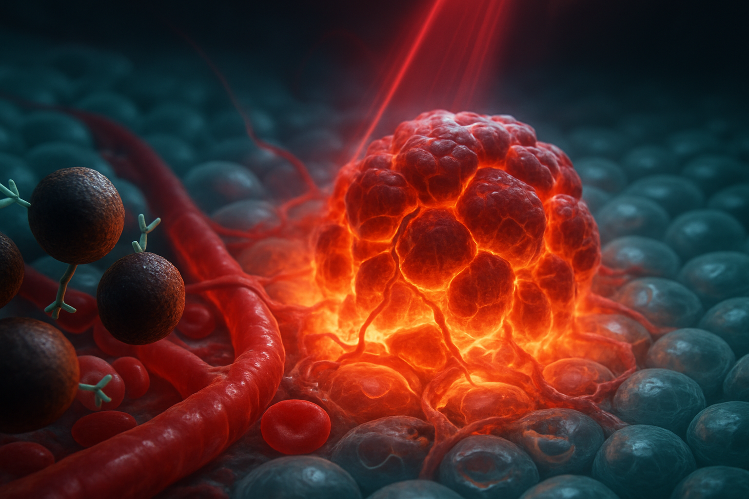
The Significance of Melanin-Coated Nanoparticles
Cancer hyperthermia aims to selectively heat malignant tissues to damage or destroy tumor cells. However, the challenge lies in controlling the spatial distribution of this heat to avoid damaging surrounding healthy tissues. Photothermal therapy has emerged as a promising strategy by employing nanoparticles that act as localized heat absorbers. This method offers a potential shift from widespread thermal procedures to finely-targeted local interventions.
A melanin-coated nanoparticle comprises a central core encapsulated within a melanin-based layer. This layer can absorb optical energy, serve as an interface for chemical modifications, or fulfill both roles. Additional elements such as targeting ligands, therapeutic agents, and imaging markers can be incorporated. This multifunctional nanoparticle design aims to achieve tumor-specific localization, near-infrared (NIR) photothermal conversion, integrated imaging, and combination therapeutic capabilities.
While the conceptual framework is attractive, current evidence underscores the necessity for further validation. The vast array of existing research provides a foundation in nanoparticle technology, surface chemistry, and tumor biology, but lacks a standardized formulation of melanin-coated nanoparticles with published criteria of safety and efficacy.
The Quantum Melanin Research Foundation acknowledges the potential of merging optical physics and tumor biology as a pivotal advancement in biophysics. The success of melanin-coated nanoparticles will depend on their ability to repeatedly deliver precise thermal doses specific to tumors under realistic clinical conditions.
Exploring NIR-Activated Hyperthermia
Photon Absorption and Heat Generation
The process begins when NIR photons are absorbed by nanoparticles, exciting electrons and charge distributions. Instead of releasing energy through light emissions, these particles convert absorbed energy into heat by nonradiative processes. This heat dispersion is influenced by numerous factors such as particle concentration, absorption efficiency, and the interaction of heat with surrounding tissue. Wavelength-specific absorption, conversion efficiency, and thermal kinetics must be accurately measured for effective material characterization.
It is crucial to differentiate photothermal therapy from photodynamic therapy, where the latter relies on light-induced reactive oxygen species to cause cell damage. Incorporation of appropriate photosensitizers could eventually allow melanin-coated nanoparticles to engage in both therapies, yet each mechanism operates on distinct principles and should be separately verified.
NIR-I, NIR-II, and Tissue Penetration
Photothermal nanomedicine employs two primary optical windows: NIR-I (approximately 650–950 nm) and NIR-II (approximately 1,000–1,700 nm). These wavelengths penetrate tissues more effectively than visible light while minimizing scattering, which may allow deeper reach to tumor cells. However, successful penetration hinges on numerous variables including tumor depth, intrinsic tissue properties, and particle distribution. Therefore, both light delivery and particle placement must be carefully coordinated to optimize therapeutic outcomes.
Currently, optimizing the absorption spectrum for melanin coatings across NIR ranges remains an open design challenge. Establishing a specific, effective NIR absorption profile for melanin-coated nanoparticles, compared to existing gold nanoparticle models, represents a significant step toward functional photothermal applications.
Emphasizing Thermal Dose over Simple Temperature
In the context of cancer therapy, mild or subablative hyperthermia typically refers to temperatures ranging from 42–45 °C. Effects at this temperature range include stressing cancer cells and enhancing membrane permeability, which may augment drug penetration and immune response. Conversely, higher temperatures cause cellular destruction through irreversible damage.
The determinant of therapeutic efficacy is thus reliant upon the thermal dose—a function of temperature plus exposure duration, spatial distribution, and tissue conductivity. Potential benefits and detriments cannot be gauged by temperature alone; numerous biological and thermal factors dictate cancer cell susceptibility.
The gap in assessing melanin-specific thermal conversion efficacy emphasizes the need for well-defined testing protocols that include long-term performance and safety assessments.
Advanced Design Considerations for Melanin Coatings
The Surface Role as Biological Control Interface
The behavior of nanoparticles in physiological environments is not solely defined by their core properties but also by their surface chemistry. Colloidal stability, protein adsorption, immune system evasion, and tissue distribution are dictated by factors such as surface charge, hydrophilicity, and ligand presentation. Particles further acquire a protein corona in biological fluids, which can influence targeting efficacy and particle biodistribution.
Melanin offers a promising platform for affixing biological targeting agents, therapeutic drugs, imaging agents, or stimuli-responsive modifications. Defining these capabilities in formulated solutions under physiological and experimental conditions will be critical to in vivo success.
Mechanisms of Targeting and Delivery
Nanoparticles employ multiple targeting strategies: passive accumulation through leaky tumor vessels; active targeting through specific ligand-receptor interactions; biomimetic approaches with biological materials to navigate immunological barriers; or responsive targeting triggered by tumor microenvironment features such as acidity or hypoxia.
Smart nanoparticle systems often merge these methods, yet the uncertainty remains high for consistent tumor selectivity. Comprehensive assessment of these targeting mechanisms in melanin-specific systems is needed to establish verifiable data reflecting enhanced tumor targeting and selective heating.
Evaluating Tumor Localization for Effective Heating
Nanoparticle presence within tumors does not automatically translate to malignant-cell selectivity. Their distribution may be skewed toward vascular regions or immune cells, thereby affecting the thermal field. Validating therapeutic impact requires more than macro-level imaging; it demands meticulous tracing of particle fate, heat distribution, and assessing tumor regions affected versus spared.
Such evaluations require measuring blood circulation, organ distribution, particle localization within cellular structures, and correlating these with heat maps to assess treatment precision and systemic exposure.
From Concept to Clinical Translation
Addressing Spatial Thermal Variation
Tumors should be modeled as heterogeneous thermal landscapes rather than consistent temperature zones. Nanoparticle concentrations, light parameters, and tissue properties all contribute to localized heat distribution. Real-time, high-resolution thermographic techniques and histological analyses are necessary to visualize and measure localized therapy effects accurately.
Discrepancy Between In Vitro and In Vivo Success
Controlled lab settings simplify challenges faced within in vivo systems, where complex biological matrices, particle aggregation, and optical attenuation hinder uniform particle distribution and heat propagation. Strategies effective in vitro may falter due to these intricate in vivo environments.
Comprehensive evaluation of nanoparticle distribution patterns, thermal gradients, tumor boundaries, and collateral tissue impacts are crucial for translating lab findings into clinical effectiveness. Both moderate and higher thermal dosages must be examined to balance efficacy with safety.
Hypothetical Validation Experiment
A potential validation could involve testing varied nanoparticle formulations: uncoated, hydrophilic-coated, melanin-coated, and melanin-coated with targeting ligands. Controls without nanoparticles would establish baselines. Characterizing particle stability, absorption, conversion, and robustness within serum systems will define initial feasibility.
Within animal models, evaluating distribution and localization across significant tissue types would provide insights into which formulations achieve superior therapeutic results under standardized conditions. Comparisons would be made among formulations to ascertain the melanin-specific impact and possibly demonstrate an enhanced therapeutic index.
Integration of Therapy and Tumor-Environment Influence
Multifunctional Treatment Frameworks
Photothermal therapies are coalescing with chemotherapeutics, immunomodulatory treatments, and agent-responsive carriers. By enhancing permeability, access, and targeting, melanin-coated nanoparticles could improve complex treatment regimens. Each additional function, however, compounds fabrication and testing complexity, necessitating further evaluation of function compatibilities within true biological contexts.
Remodeling Tumor Microenvironments
The potential for nanoparticles to remodel the tumor microenvironment—impacting vascular structures, immune profiles, and extracellular matrices—is transforming therapeutic strategies. Nevertheless, these effects may include both cancer-targeted and broad inflammatory responses. Monitoring and understanding these varied interactions is central to harnessing the full potential of this technology.
Potential for a Multifunctional Therapeutic Solution
Envisioning melanin coatings as truly multifunctional interfaces is speculative but conceivable. Integrating optical, targeting, and drug-delivery properties within one construct would simplify current layered approaches, provided rigorous compatibility and stability tests show promising results.
Immune Priming Beyond Hyperthermia
Strategically using subablative hyperthermia to prime immune responses rather than focus on tumor ablation poses a novel avenue for cancer treatment. As immune interactions can vary broadly, deep immunological assessments and longitudinal studies would be needed, particularly concerning melanin's potential contributions beyond being a thermal agent.
Translational Pathway for Clinical Application
Navigating this path requires thorough preclinical validation, encompassing materials characterization, in vivo modeling, safety, and efficacy standardization. Tracking biodistribution, clearance, immune activation, and systemic impacts represents the groundwork necessary to move from investigational use to clinical trials.
Key Takeaways
Melanin-coated nanoparticles offer a potential convergence of NIR photothermal treatments, targeted delivery systems, imaging capabilities, and multifaceted therapeutic function. Verification of their effectiveness will rest on rigorous trials assessing their capabilities against established benchmarks.
In the mild-hyperthermia range of 42–45 °C, outcomes will largely depend on comprehensive considerations spanning thermal dosing, exposure durations, particle localization, and tissue-specific responses. Tumor homing alone is insufficient for malignant specificity—detailed analysis of microenvironmental interactions is paramount.
Presently, definitive melanin-specific photothermal efficiency and therapeutic data is lacking, prioritizing targeted validation experiments against existing standards. Thus, while promising, melanin-coated nanoparticles should be considered as innovative research platforms pending further development and scrutiny.
Their potential lies in unifying conventional therapies with nanotechnological advancements—an interdisciplinary approach that could redefine therapeutic practices pending proof of efficacy and reproducibility.
Related Research
- Proton Tunneling in Melanin: Implications for Biological Energy Storage and Transfer
- Could Melanin Enable Photosynthesis-Like Energy Production in Human Cells?
- The Evolutionary Significance of Melanin: Why Nature’s Most Ancient Pigment Persists Across the Tree of Life
References
- Yihan Yao, Yunxiang Zhou, Lihong Liu. "Nanoparticle-Based Drug Delivery in Cancer Therapy and Its Role in Overcoming Drug Resistance." Frontiers in Molecular Biosciences. 2020. doi:10.3389/fmolb.2020.00193
- Leming Sun, Hongmei Liu, Yanqi Ye. "Smart nanoparticles for cancer therapy." Signal Transduction and Targeted Therapy. 2023. doi:10.1038/s41392-023-01642-x
- Jeremy B. Vines, Jee-Hyun Yoon, Na-Eun Ryu. "Gold Nanoparticles for Photothermal Cancer Therapy." Frontiers in Chemistry. 2019. doi:10.3389/fchem.2019.00167
- Gennaro Sanità, Barbara Carrese, Annalisa Lamberti. "Nanoparticle Surface Functionalization: How to Improve Biocompatibility and Cellular Internalization." Frontiers in Molecular Biosciences. 2020. doi:10.3389/fmolb.2020.587012
- Li Chen, Weiqi Hong, Wenyan Ren. "Recent progress in targeted delivery vectors based on biomimetic nanoparticles." Signal Transduction and Targeted Therapy. 2021. doi:10.1038/s41392-021-00631-2
- Qiang Lü, Dongquan Kou, Shenghan Lou. "Nanoparticles in tumor microenvironment remodeling and cancer immunotherapy." Journal of Hematology & Oncology. 2024. doi:10.1186/s13045-024-01535-8
- Di Gao, X. Guo, Xingcai Zhang. "Multifunctional phototheranostic nanomedicine for cancer imaging and treatment." Materials Today Bio. 2019. doi:10.1016/j.mtbio.2019.100035
- Xiulin Yi, Qiu‐Yi Duan, Fu‐Gen Wu. "Low-Temperature Photothermal Therapy: Strategies and Applications." Research. 2021. doi:10.34133/2021/9816594