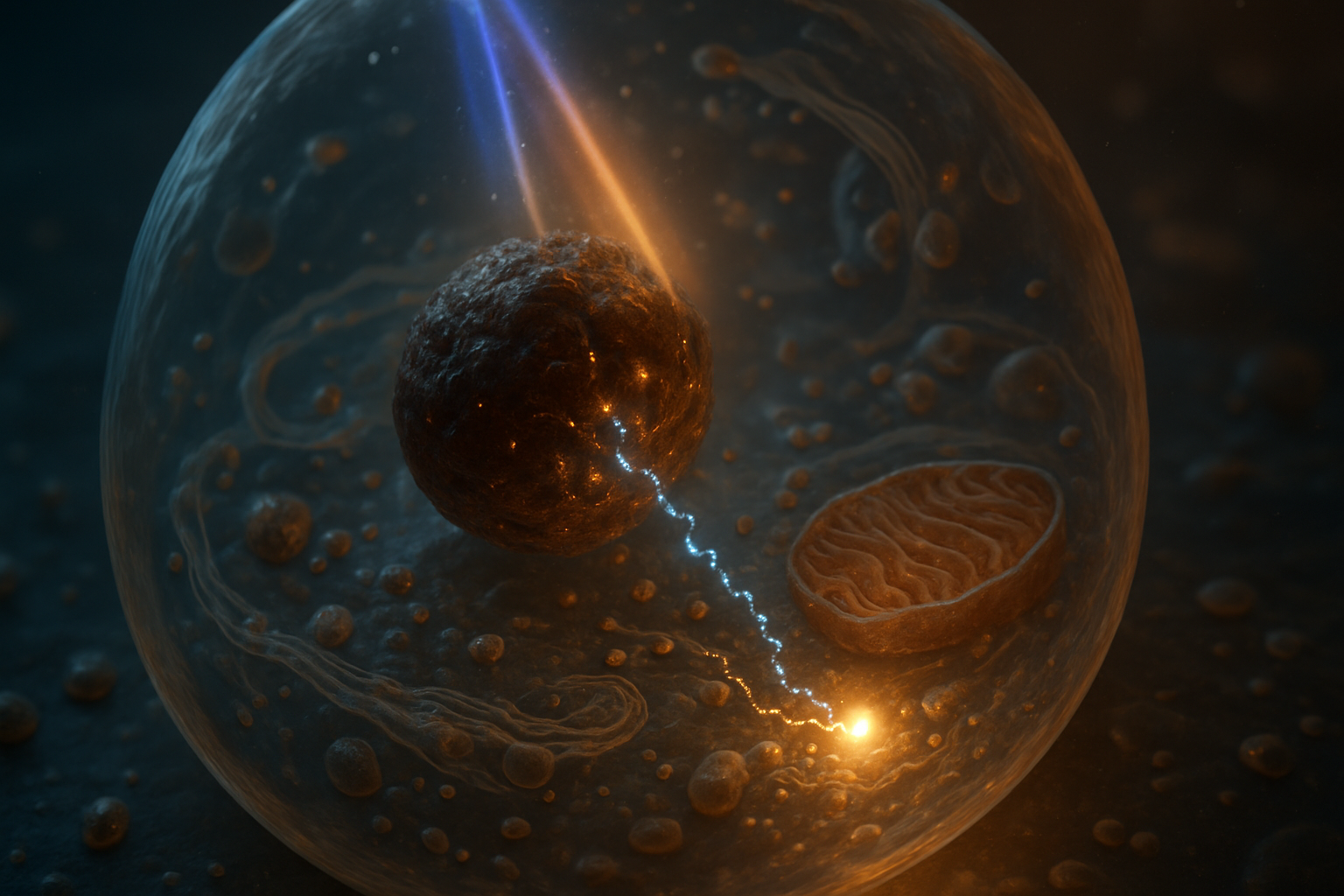
Introduction: Transforming Melanin from a Light-Absorbing Pigment to a Cellular Power Source
Melanin is widely recognized as the pigment responsible for absorbing light in biological systems. In skin, hair, the retinal pigment epithelium, the inner ear, and parts of the brain, it is best known for limiting photochemical injury by absorbing ultraviolet and visible radiation. However, a more ambitious inquiry follows from melanin’s unusual electronic and chemical properties: Could this pigment transform some fraction of absorbed light into metabolically useful energy within human cells?
“Photosynthesis-like” can refer to several different levels of function. At the weakest level, melanin might alter cellular redox chemistry after illumination. At a more substantial level, it might transfer photoinduced electrons to an acceptor, reduce a metabolic cofactor, or influence membrane charge. The strongest claim would be that melanin participates in a complete energy-conserving pathway that generates a proton-motive force, produces ATP, or drives a catalytic process such as water oxidation.
These possibilities must be distinguished carefully. A pigment can absorb photons without storing their energy in a useful chemical form. It can also produce heat, transient radicals, or electrical signals without creating a sustainable source of cellular work. Photosynthesis is not simply “light absorption.” It is a coordinated system involving:
- photon capture;
- charge separation;
- directional electron transfer;
- suppression of charge recombination;
- coupling to proton movement or another energy-storage mechanism;
- regeneration of the initial redox state; and
- connection to metabolism.
In oxygenic photosynthesis, these functions are distributed across organized pigment–protein complexes and membranes. Chlorophyll absorbs photons in a carefully structured environment, reaction-center cofactors separate charge, electron carriers pass electrons in a defined direction, and membrane-bound complexes establish a proton gradient. ATP synthase then converts that electrochemical gradient into ATP. A melanin-containing human cell would need to reproduce at least some of these functions despite lacking chloroplasts, photosynthetic reaction centers, and dedicated photosynthetic membranes.
Some researchers have proposed that melanin can convert light into heat, electrical charge, or altered redox states. Other proposals have suggested that melanin might participate in water-splitting chemistry or provide an alternative energy pathway in human tissue. These ideas are scientifically interesting, but they remain hypotheses unless supported by quantitative measurements demonstrating energy conservation and metabolic coupling.
The relevant question is therefore not merely whether melanin responds to light. It plainly does. The central question is whether the response can produce a sustained, directional, and biologically useful flow of energy that exceeds the energetic and oxidative costs of the system.
Understanding Melanin’s Photophysics: Established Knowledge and New Proposals
Melanin: A Versatile Light Absorber
Melanin is not a single, uniform compound. The principal forms in humans include eumelanin, which is brown to black, and pheomelanin, which is reddish to yellow and contains sulfur-bearing units. Neuromelanin is a chemically and structurally distinct, organelle-associated pigment found in specific neuronal populations and is thought to contain both melanin-like polymers and bound metals, lipids, and other cellular components.
Eumelanin is commonly described as a heterogeneous polymer or supramolecular assembly derived largely from indole-based units such as 5,6-dihydroxyindole and its oxidized derivatives. Its electronic behavior reflects a distribution of chemically different subunits, oxidation states, protonation states, and aggregate structures rather than one sharply defined molecular transition. This helps explain one of melanin’s most characteristic optical features: broad, relatively featureless absorption that generally decreases as wavelength increases.
Broad absorption is useful for photoprotection because it allows melanin to intercept radiation over a wide range of wavelengths. It does not, however, demonstrate high photochemical efficiency. The crucial quantities would include:
- the absorption coefficient at each relevant wavelength;
- the fraction of incident light reaching the melanin;
- the quantum yield of charge separation;
- the lifetime of separated charges;
- the proportion of excitation energy retained rather than dissipated;
- the rate of productive electron transfer; and
- the fraction ultimately converted into ATP or another stable chemical product.
These measurements vary substantially depending on whether the sample is synthetic melanin, isolated natural melanin, melanosomes, a protein-associated preparation, or pigment embedded in a living cell. Hydration, pH, oxygen concentration, metal binding, particle size, aggregation, and oxidation state can all change the apparent optical and electrochemical response.
A useful distinction is between optical absorption and usable photon capture. If a pigment layer absorbs 90% of incoming light but converts 99.9% of that energy into heat, its energy-conversion efficiency is still extremely low. Conversely, a pigment that absorbs less light might be more useful if it directs a larger fraction into long-lived charge separation. Photosynthetic organisms solve this problem through molecular organization; melanin’s disordered structure may be advantageous for broadband protection but disadvantageous for directional energy transfer.
A simple photon-energy calculation illustrates the scale involved. The energy of one photon is:
[ E = \frac{hc}{\lambda} ]
where (h) is Planck’s constant, (c) is the speed of light, and (\lambda) is wavelength. A photon at 500 nanometers carries approximately (3.97 \times 10^{-19}) joules, or about 239 kilojoules per mole of photons. A 700-nanometer photon carries approximately 171 kilojoules per mole. This is enough energy, in principle, to drive many biochemical reactions, but only if the energy is captured, stabilized, and transferred before it is lost.
Energy Dissipation in Melanin as Heat
Melanin is often described as converting absorbed light into heat. This is consistent with a photoprotective role. Excited electronic states can relax through internal conversion, vibrational motion, proton transfer, and other nonradiative pathways. The energy eventually appears as molecular motion and raises the local temperature by a small amount.
For photoprotection, rapid dissipation is beneficial. A long-lived excited state could instead transfer energy to oxygen and generate singlet oxygen or other reactive oxygen species. Melanin’s low fluorescence yield and strong nonradiative relaxation are therefore commonly interpreted as evidence that much of the absorbed excitation energy is safely dissipated.
A rough physical estimate shows why bulk heating alone is unlikely to supply a major ATP source. Suppose a small tissue volume absorbs 1 milliwatt of optical power for 1 second. It receives 1 millijoule of energy. If that energy is distributed through 1 milligram of water-equivalent tissue, the temperature rise would be approximately:
[ \Delta T = \frac{Q}{mc_p} ]
Using (c_p \approx 4.2\ \text{J g}^{-1}\text{K}^{-1}), the temperature increase would be about 0.24 kelvin if heat were initially confined to that mass. In a living tissue, diffusion and blood flow would rapidly spread the heat. More importantly, heat is not automatically convertible into ATP. A cell would need a heat engine operating across a temperature gradient, and biological temperature differences are generally too small and too transient to support an efficient ATP-producing mechanism.
This does not mean that heat is biologically irrelevant. Local heating can influence membrane properties, enzyme rates, blood flow, and stress responses. Melanin-containing materials are also being investigated for photothermal therapy, in which heat is intentionally used to damage tumor cells. Photothermal utility, however, is conceptually different from photochemical energy conservation.
The key experimental question is therefore the energy partition:
[ E_{\text{absorbed}} = E_{\text{heat}} + E_{\text{fluorescence}} + E_{\text{chemical}} + E_{\text{electrical}} ]
In a living cell, these terms are difficult to measure independently. A claim of energy production would require showing that the chemical or electrochemical term is both reproducible and large enough to influence metabolism.
Light-Induced Charge and Redox Changes in Melanin
Melanin can exhibit electrically and electrochemically interesting behavior. Depending on its hydration and chemical environment, it may support proton conduction, ionic movement, electron hopping, or mixed ionic–electronic transport. Changes in pH, humidity, metal binding, and oxidation state can alter its conductivity. These properties have motivated research into melanin-based sensors, bioelectronic materials, and organic electrochemical devices.
Illumination may produce measurable changes in current, open-circuit potential, impedance, or surface charge. Such responses could arise from several mechanisms:
- excitation of localized electronic states;
- photoinduced redistribution of electrons among quinone and hydroquinone groups;
- movement of protons coupled to redox reactions;
- changes in hydration or dielectric properties;
- release or capture of metal ions;
- photothermal changes in conductivity; or
- photooxidation involving oxygen or other acceptors.
A current measured through a melanin film does not by itself establish biological energy production. Electrical current can result from an external voltage, electrode reactions, ionic gradients, or a transient discharge of stored charge. To establish a productive photochemical cycle, researchers would need to measure the charge generated per incident photon, determine whether the charge persists long enough to be transferred, identify the electron donor and acceptor, and demonstrate repeated cycling without progressive chemical degradation.
For example, if illumination produces a photocurrent of 1 nanoampere for 100 seconds, the total transferred charge is:
[ Q = It = (1 \times 10^{-9}\ \text{A})(100\ \text{s}) = 1 \times 10^{-7}\ \text{C} ]
This corresponds to approximately (6.2 \times 10^{11}) elementary charges. That number may sound large, but its energetic significance depends on the potential difference and whether the current is sustained by light or by depletion of a finite redox reservoir. At a potential of 0.2 volts, the electrical work is approximately (2 \times 10^{-8}) joules. That is a measurable laboratory signal, but it must be compared with incident optical energy and with the energy demands of the cell.
The duration of charge separation is equally important. A charge that recombines in picoseconds is unlikely to drive a distant metabolic reaction unless a precisely positioned acceptor is present. A charge separated for microseconds, milliseconds, or longer has a greater opportunity for productive transfer, but lifetime alone still does not establish useful energy conversion. The direction, yield, and chemical reversibility of the transfer must also be demonstrated.
What Photosynthesis Entails: From Capturing Photons to Chemical Tasks
Photosynthesis is an intrinsic system starting with photon absorption, passing through a series of electron transfers, and ending with the storage of chemical energy. Core elements include capturing photons, creating charge separation, electron transfer, proton movement, and energy storage in forms such as ATP and NADPH.
In oxygenic photosynthesis, the pathway is spatially organized within a membrane. Photosystem II extracts electrons from water and releases oxygen. The cytochrome (b_6f) complex contributes to proton translocation. Photosystem I generates highly reducing electrons that ultimately help reduce NADP(^+) to NADPH. ATP synthase uses the proton-motive force generated across the membrane.
The electrochemical free energy available from a proton gradient can be represented approximately as:
[ \Delta p = \Delta \psi - \frac{2.303RT}{F}\Delta pH ]
where (\Delta \psi) is the electrical potential difference, (R) is the gas constant, (T) is absolute temperature, and (F) is Faraday’s constant. Mitochondrial ATP synthase commonly operates with a proton-motive force on the order of roughly 150–200 millivolts, although the exact value varies with tissue, metabolic state, and experimental conditions.
ATP synthesis is not free. Depending on the organism and enzyme, approximately three to four protons may be required per ATP synthesized through the catalytic motor, with additional energetic costs associated with transporting phosphate, ADP, ATP, and other metabolites. A photosynthesis-like melanin system would therefore need to move a substantial number of protons in a controlled direction and across a defined membrane barrier.
Water Splitting: A Challenging Endeavor
The hypothesis that melanin could drive water splitting analogous to chlorophyll is especially demanding. Water oxidation is not simply the release of oxygen from illuminated water. In oxygenic photosynthesis, the oxygen-evolving complex of photosystem II accumulates multiple oxidizing equivalents through a catalytic cycle. Four electrons are removed from two water molecules to form one oxygen molecule:
[ 2H_2O \rightarrow O_2 + 4H^+ + 4e^- ]
This reaction requires a sufficiently strong oxidant and a mechanism capable of storing multiple redox equivalents without destructive side reactions. The standard thermodynamic potential for water oxidation is approximately (+1.23) volts under standard conditions, with the actual potential depending on pH, oxygen pressure, and substrate activities. At neutral pH and physiological conditions, the energetic requirement remains substantial.
A credible claim of melanin-driven water oxidation would need to show all of the following:
- oxygen production increases specifically with illumination;
- the amount of oxygen follows the expected four-electron stoichiometry;
- the oxygen originates from water rather than dissolved oxygen, peroxide decomposition, or another contaminant;
- the process is catalytic and repeatable;
- the pigment remains chemically intact or is demonstrably regenerated;
- appropriate dark, oxygen-free, heat-matched, and pigment-free controls fail to produce the same signal; and
- isotope labeling, such as (^{18})O-enriched water, confirms the source of the evolved oxygen.
A dissolved-oxygen electrode can detect changes in oxygen concentration, but such measurements are vulnerable to artifacts. Air equilibration, photosensitive impurities, electrode heating, peroxide chemistry, and microbial contamination can all produce apparent oxygen evolution. Isotope-ratio mass spectrometry or membrane-inlet mass spectrometry would provide stronger evidence than oxygen readings alone.
The reduction half-reaction must also be identified. If water supplies electrons, those electrons must reduce a defined acceptor, such as NADP(^+), quinone, a metal center, or another redox mediator. Measuring oxygen without accounting for the corresponding electron sink is incomplete redox bookkeeping.
Achieving Photosynthetic Function
For melanin to mimic even part of chlorophyll-based photosynthesis, several conditions must be satisfied:
- Dependence on light: The effect should increase with photon dose within a defined range and show a reproducible action spectrum.
- Energy conservation: The absorbed optical energy should appear in a stable chemical or electrochemical product rather than only as heat.
- Charge balance: Oxidation and reduction products must be accounted for quantitatively.
- Catalytic turnover: The pigment or system should operate repeatedly rather than being consumed in a single photochemical event.
- Directional transfer: Electrons or protons must move toward defined acceptors or across a defined membrane.
- Metabolic coupling: The output must measurably affect ATP, NAD(P)H, carbon flux, ion transport, or another cellular function.
- Physiological relevance: The effect must remain after accounting for heating, stress responses, altered oxygen availability, and experimental artifacts.
A worked example helps define the scale. Suppose a cell receives 1 picowatt of absorbed optical power for 1 second. This is (10^{-12}) joules, or approximately (2.5 \times 10^{-14}) moles of 500-nanometer photons if every photon were absorbed. If 1% of those photons generated a productive electron transfer, the cell would receive roughly (2.5 \times 10^{-16}) moles of electrons per second. That is potentially measurable with sensitive techniques, but it does not automatically translate into a meaningful ATP supply. The electrons must be connected to a pathway, and the energy per electron must be sufficient to drive an energetically uphill reaction.
If a cell instead absorbed 100 picowatts and converted 1% of the input into biochemical work, the usable power would be 1 picowatt. This could be relevant in a very small or energy-limited compartment, but it would still need to be compared with basal cellular power consumption, which varies widely by cell type and physiological state. The example also shows why both photon flux and conversion efficiency must be measured rather than inferred from pigmentation.
The Limits of Charge Generation
Free charges can dissipate through recombination, trapping, electrode contact, oxygen reactions, or random redox chemistry. A cell requires a mechanism to direct them. In a membrane, that directionality may be established by the orientation of redox proteins and the physical separation of electron donors and acceptors. In an unstructured melanin aggregate, electrons may instead move through a distribution of localized states with substantial back-reaction.
A useful distinction is between:
- stored charge, which may be released later;
- transient photocurrent, which reflects a short-lived redistribution;
- redox turnover, which involves repeated oxidation and reduction; and
- energy-conserving electron transfer, which produces a usable gradient or chemical product.
Only the latter two are candidates for metabolic coupling, and even redox turnover may simply generate oxidative stress if it is not integrated into a controlled pathway.
ATP synthase illustrates the architectural requirement. The enzyme does not use free electrons directly to make ATP. It uses rotation driven by proton flow through the (F_o) portion of the complex. Protons must be accumulated on one side of a membrane, the membrane must limit passive leakage, and the gradient must be sufficiently stable to drive conformational changes in the catalytic (F_1) portion. Melanin-induced charge changes in the cytosol, nucleus, or a disordered organelle would not automatically create the required transmembrane proton-motive force.
The Feasibility of Integrating a Melanin-Based Energy System in Human Cells
Possible Outcomes of Melanin Light Interaction
Human cells lack chloroplasts, photosynthetic reaction centers, and the specialized membranes used for light-driven proton pumping. They do possess mitochondria, redox enzymes, proton gradients, antioxidant systems, and extensive membrane structures. In principle, these existing components could provide partial support for a light-responsive pathway, but they could also create competing sinks and sources of reactive chemistry.
A viable melanin–light system might produce one or more of the following outputs:
- increased ATP concentration or ATP production rate;
- an increase in mitochondrial or other membrane potential;
- a measurable proton gradient;
- reduction of NADP(^+), NAD(^+), quinones, or another defined cofactor;
- altered oxygen consumption consistent with useful electron input;
- increased carbon fixation or biosynthetic flux; or
- improved survival under a defined energetic stress.
Each output requires careful interpretation. An increase in ATP after illumination could result from calcium signaling, stress-induced metabolic reprogramming, increased glycolysis, mitochondrial uncoupling responses, or temperature changes rather than direct photon-to-ATP conversion. Likewise, a change in membrane potential could reflect ion-channel activity or photothermal membrane perturbation rather than proton pumping.
Photon availability imposes another constraint. Sunlight at Earth’s surface can approach approximately 1,000 watts per square meter under strong midday conditions, but the biologically relevant irradiance depends on wavelength, cloud cover, tissue depth, pigmentation, and exposure geometry. Human skin receives light at the surface, whereas cells deep in tissue receive far less, especially in the ultraviolet and blue regions. Melanin absorbs strongly, which protects underlying cells but also limits the penetration of light to deeper pigment-containing compartments.
A second constraint is area. A photosynthetic leaf presents a large light-collecting surface relative to its metabolic machinery. A single human cell has a microscopic area. Even if it absorbed light efficiently, the total photon power available to it would remain limited. For example, a cell with an effective projected area of (100\ \mu\text{m}^2) exposed to (100\ \text{W m}^{-2}) of usable light would intercept only:
[ P = IA = (100\ \text{W m}^{-2})(100 \times 10^{-12}\ \text{m}^2) = 10^{-8}\ \text{W} ]
or 10 nanowatts before accounting for shading, reflection, incomplete absorption, and conversion losses. A 1% conversion efficiency would yield 0.1 nanowatt. This is not necessarily negligible, but it is far below the power demands of an actively functioning cell if interpreted as a universal substitute for mitochondrial metabolism.
Weighing Biological Trade-offs
Any system pairing melanin with photochemical processes must address biological safety. Illumination may produce reactive oxygen species through several routes, including photooxidation of melanin, reduction of oxygen to superoxide, formation of hydrogen peroxide, and generation of singlet oxygen. Pheomelanin has often been associated with greater photochemical oxidative stress than eumelanin under some conditions, although the outcome depends strongly on the molecular environment.
Redox-active melanin could also bind and redistribute metals such as iron or copper. This might buffer toxic metals, as proposed for some forms of neuromelanin, but it could also promote localized Fenton-type chemistry if reducing equivalents and peroxide are present. A system that generates a small amount of ATP while increasing lipid peroxidation, DNA damage, mitochondrial injury, or inflammatory signaling would not represent a beneficial energy technology.
The net energetic balance can be expressed conceptually as:
[ P_{\text{net}} = P_{\text{captured and conserved}}
P_{\text{repair}}
P_{\text{detoxification}}
P_{\text{system maintenance}} ]
Repair costs include antioxidant regeneration, removal of oxidized proteins, membrane repair, DNA repair, and replacement of damaged organelles. A photochemical pathway would also require proteins, cofactors, structural organization, and regulatory mechanisms. These support costs could exceed the energy harvested.
Safety testing would therefore need to measure more than ATP. Important endpoints would include hydrogen peroxide, superoxide, singlet oxygen, glutathione redox state, lipid peroxidation products, mitochondrial membrane potential, oxygen consumption, calcium signaling, DNA damage markers, and cell viability. Illumination should be matched for total energy and temperature, with dark controls, pigment-free controls, antioxidant controls, and wavelength-specific controls.
What If? Imagining Hypothetical Human Photosynthesis Scenarios
Scenario 1: Predominantly as a Redox Mediator
The least ambitious scenario is that melanin acts as a light-responsive redox mediator. Illumination might alter the oxidation state of quinone-like groups or change electron transfer between melanin, oxygen, metal ions, and cellular cofactors.
In this model, melanin would not make ATP directly. Instead, it might influence existing pathways—for example, by donating electrons to a defined acceptor or buffering redox fluctuations. A successful demonstration would require:
- an action spectrum showing wavelength-dependent activity;
- identification of the oxidized and reduced melanin states;
- a defined electron donor and acceptor;
- quantification of charge transfer per photon;
- repeated cycles of oxidation and reduction;
- minimal pigment degradation; and
- direct evidence that the altered redox flow changes a cellular metabolic reaction.
A worked assay could combine purified melanin or melanosomes with a known redox mediator such as a quinone analogue, NAD(^+)/NADH, or a protein electron acceptor. Researchers could measure absorbance changes, electron paramagnetic resonance signals, transient absorption, and oxygen consumption under controlled illumination. If illumination reduced the acceptor and the reduced acceptor subsequently drove a defined enzyme reaction, this would demonstrate a real photoredox function. It would still not prove ATP generation, but it would establish a mechanistic intermediate.
Scenario 2: Creating a Proton-Pumping System
A more intricate concept involves assembling melanin into a structural component of a proton-pumping mechanism that yields ATP. This would require more than photoinduced charge. Melanin would need to participate in a cycle that moves protons across a membrane or changes proton permeability in a directionally controlled way.
The minimum architecture would include:
- a light-responsive redox or proton-transfer element;
- a membrane or vesicle that separates two aqueous compartments;
- a mechanism that prevents immediate proton equilibration;
- a proton-conducting path coupled to the photochemical cycle; and
- ATP synthase positioned so that proton flow drives ATP formation.
An experimentally tractable first step would be a reconstituted liposome containing melanin-associated material and ATP synthase. The vesicles would be loaded with a pH-sensitive fluorescent dye, and illumination would be monitored with simultaneous measurements of intravesicular pH, membrane potential, and ATP production. A valid result would require a light-dependent proton gradient that disappears when the pigment, membrane, or ATP synthase is omitted.
The expected energy scale can be estimated. At room temperature, a proton-motive force of approximately 180 millivolts corresponds to roughly 17 kilojoules per mole of protons for the electrical component alone. If three to four protons are required per ATP, the gradient must provide enough free energy to exceed the approximately 30–50 kilojoules per mole free-energy cost of ATP synthesis under cellular conditions, including transport losses. A small voltage signal in a planar film would not necessarily meet this threshold.
In human cells, the problem becomes more difficult because endogenous membranes are already tightly regulated. Adding a light-responsive proton conductor could depolarize mitochondria, acidify organelles, or disrupt lysosomal and endosomal function. The system would need precise localization and gating to avoid becoming an uncoupler rather than an energy converter.
Scenario 3: Achieving Water Oxidation
At the most ambitious level, melanin would facilitate water oxidation and transfer the resulting electrons into productive metabolism. This would require a catalytic mechanism capable of accumulating four oxidizing equivalents, releasing oxygen with the correct stoichiometry, and preventing destructive oxidation of the pigment and surrounding biomolecules.
Evidence would need to combine several measurements:
- oxygen evolution under monochromatic or defined-spectrum illumination;
- (^{18})O labeling to establish water as the oxygen source;
- measurement of the corresponding reduced product;
- quantification of proton release;
- action-spectrum and intensity-dependence data;
- repeated turnover over many cycles;
- controls excluding peroxide decomposition and dissolved oxygen artifacts; and
- structural or spectroscopic identification of the catalytic redox center.
Even if oxygen evolution were confirmed in vitro, integrating it into human cells would introduce substantial hazards. Oxygen production near reduced metal centers and organic radicals could increase oxidative stress. The electrons generated from water oxidation would also need a defined destination. Without a coupled reductive pathway, water splitting would represent chemically expensive oxidation rather than useful energy production.
Hypothetical Experiment Framework
A rigorous experimental program could proceed from simple systems to living cells.
First, characterize the material. Researchers would determine melanin composition, oxidation state, hydration, particle size, metal content, absorption coefficient, and electrochemical properties. Synthetic eumelanin should not be assumed to behave identically to natural melanosomes or neuromelanin.
Second, measure photophysical events. Ultrafast spectroscopy could determine the timescale of excited-state relaxation and charge separation. Transient absorption, time-resolved electron paramagnetic resonance, and photocurrent measurements could identify persistent radicals or mobile charges. Integrating the signal over time would estimate the number of charges generated per incident photon.
Third, test defined chemical coupling. Melanin could be combined with purified electron acceptors, proton-sensitive probes, redox enzymes, or membrane vesicles. Controls should include heat-matched dark samples, inert carbon particles with similar optical absorption, antioxidant supplementation, oxygen-free conditions, and altered pH.
Fourth, test energy conservation. In vesicles, investigators could measure light-dependent proton gradients and ATP synthesis. In cell-free extracts, they could test NAD(P)H production, quinone reduction, or stimulation of an enzyme reaction. The critical comparison would be energy output versus absorbed optical input.
Fifth, test living cells. Melanized and non-melanized cells would be compared under identical illumination, temperature, oxygen, and nutrient conditions. Measurements could include ATP/ADP ratio, NADH/NAD(^+), NADPH/NADP(^+), oxygen consumption, extracellular acidification, mitochondrial potential, isotope-traced carbon flux, and reactive oxygen species.
A particularly important control would be to separate direct photochemistry from secondary biological responses. If illumination changes ATP only after several minutes and the effect is blocked by inhibitors of calcium signaling or stress kinases, the result may reflect signaling rather than photon energy capture. By contrast, a direct energy-conversion mechanism should produce an immediate, dose-dependent, wavelength-dependent change with a quantitatively traceable electron or proton flux.
Long-term experiments would also be necessary. A pigment might produce a short-lived signal during the first few illumination cycles but become oxidized, photobleached, or chemically altered. Productive photosynthetic systems are catalytic and regenerative; transient photoreactivity is not enough.
Concluding Thoughts
Melanin interacts with light in scientifically significant ways. It absorbs across broad spectral ranges, dissipates excitation energy efficiently, can participate in complex proton- and electron-transfer chemistry, and may generate measurable electrical or redox responses under some conditions. These properties make it a compelling subject for biophysics, bioelectronics, photomedicine, and cellular redox research.
However, the distance between light absorption and photosynthesis-like energy production is substantial. A genuine cellular energy pathway would require organized charge separation, directional transfer, proton or chemical energy storage, regeneration of the pigment’s active state, and coupling to ATP synthesis or another metabolic process. Human cells do not naturally possess the reaction centers or membrane architecture that make oxygenic photosynthesis efficient.
The most plausible near-term possibility is not that melanin turns human cells into plants, but that it may act as a light-sensitive redox or electronic material that modifies existing cellular chemistry. Whether such modification is beneficial depends on the magnitude of the effect, its reproducibility, its energetic efficiency, and its oxidative cost. A small increase in redox activity could be useful in a carefully engineered device, while the same activity in a living cell could generate damaging radicals.
Progress therefore depends on quantitative evidence. Researchers must distinguish absorbed photons from conserved energy, transient charge from sustained electron transfer, oxygen detection from water oxidation, and altered ATP levels from direct photon-to-ATP conversion. Measurements of quantum yield, charge lifetime, proton gradients, oxygen isotope labeling, ATP production, and oxidative damage should be integrated rather than considered separately.
The QMRF views melanin-based bioenergetics as a frontier question in biophysics. The hypothesis is not established, but it is testable. Carefully designed experiments could determine whether melanin is primarily a photoprotective dissipater, a redox mediator, a bioelectronic material, or—under specially engineered conditions—a component of a more elaborate energy-conversion system. Intellectual curiosity is warranted, but the decisive standard must remain reproducible, mechanistically resolved, and energetically meaningful evidence.