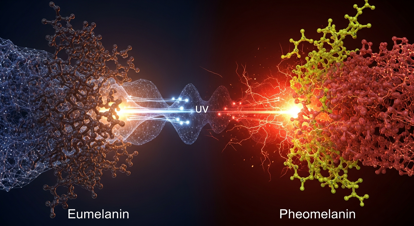
While traditionally viewed simply as biological colorants, the two primary forms of human melanin are fundamentally distinct macromolecular systems. Their structural disparities dictate completely opposite biophysical behaviors—one acts as an exceptionally efficient energy dissipator, while the other serves as a latent generator of oxidative stress.
In 2012, a landmark study out of Massachusetts General Hospital sent shockwaves through the dermatological and cancer biology communities. Researchers investigating the unusually high rates of melanoma in red-haired, fair-skinned individuals made a startling discovery: the elevated cancer risk was not merely a result of this phenotype offering less UV protection. Instead, the pigment itself—pheomelanin—was actively driving carcinogenesis in the complete absence of ultraviolet light. This finding fundamentally disrupted the classical view of melanin as an inert biological parasol. It forced a paradigm shift in biophysics, revealing that the melanin polymer is an active participant in cellular redox chemistry, capable of both safeguarding the genome and dismantling it.
To understand how a single class of biomacromolecules can exhibit such a profound biophysical schism, one must look past the macroscopic trait of pigmentation and examine the quantum and molecular architecture of the polymers themselves. At the Quantum Melanin Research Foundation (QMRF), we approach melanins not as static colors, but as dynamic, solid-state biopolymeric networks. By dissecting the structural disparities between eumelanin and pheomelanin, we can decode the physical mechanisms that govern their contrasting roles in photoprotection, bioelectricity, and cellular survival.
The Eumelanin Shield: Broadband Absorption and Ultrafast Dissipation
The dark brown-to-black pigment known as eumelanin is widely considered one of nature’s most robust photoprotective agents. Its protective capacity is not merely a function of its dark color, but is instead an emergent property of its highly cross-linked molecular structure. Eumelanin is an amorphous semiconductor assembled primarily from the oxidative polymerization of two monomeric precursors: 5,6-dihydroxyindole (DHI) and 5,6-dihydroxyindole-2-carboxylic acid (DHICA).
The integration of these DHI and DHICA monomers creates what biophysicists call a "chemical disorder" model. Because the monomers polymerize in a variety of randomized planar oligomers—each with slightly different electronic properties and energy states—eumelanin exhibits a completely unstructured, broadband absorption spectrum. It absorbs photons across the UV and visible light spectrum continuously, with an optical bandgap of approximately 1.85 electron volts (eV).
However, absorbing a highly energetic UV photon is only half the battle; the molecule must safely dispose of that energy before it can damage surrounding cellular structures. Eumelanin achieves this through a quantum mechanical process known as internal conversion. When a photon strikes a eumelanin oligomer, the molecule transitions to an excited electronic state. Within femtoseconds (quadrillionths of a second), the polymer funnels this excitation energy into molecular vibrations.
Think of eumelanin as a molecular shock absorber. It takes the violent, high-frequency impact of ultraviolet radiation and disperses it as low-frequency heat (phonons) into the surrounding solvent. Studies by physical chemists, notably the work of Paul Meredith and Tadeusz Sarna, have demonstrated that eumelanin’s non-radiative relaxation efficiency exceeds 99.9%. By instantly thermally dissipating absorbed energy, eumelanin prevents the formation of long-lived excited states that could otherwise trigger erratic, damaging photochemistry in the cell.
Pheomelanin and the Benzothiazine Trap
Pheomelanin, the yellow-to-red pigment dominant in individuals with red hair and fair skin, represents a dramatic evolutionary and structural divergence. While both melanins begin with the oxidation of the amino acid tyrosine, pheomelanogenesis takes a sharp detour when the intermediate dopaquinone reacts with cysteine, a sulfur-containing amino acid. This reaction produces cysteinyldopa, which subsequently cyclizes to form benzothiazine and benzothiazole units.
The inclusion of sulfur and the resulting benzothiazine geometry fundamentally sabotage the highly conjugated, planar networks that give eumelanin its protective prowess. Pheomelanin polymers are more disjointed, possess a lower molecular weight, and exhibit a vastly different electronic landscape.
Because of this fractured architecture, pheomelanin fails to execute efficient internal conversion. When pheomelanin absorbs UV radiation, the excitation energy becomes "trapped" in longer-lived triplet states. This represents a biophysical hazard. In this excited state, pheomelanin acts as a photosensitizer. Instead of dissipating the energy as heat, the excited polymer interacts with molecular oxygen ($O_2$) within the cellular environment. Through direct electron transfer, pheomelanin reduces oxygen to generate highly destructive reactive oxygen species (ROS), including superoxide anions and singlet oxygen.
Under UV radiation, the very molecule synthesized to protect the cell turns against it, initiating cascades of lipid peroxidation, protein damage, and DNA mutations. Pheomelanin is not merely a weaker shield than eumelanin; under the stress of high-energy photons, it becomes an active weapon.
The Redox Tug-of-War: From Antioxidant to Pro-Oxidant
The biophysical consequences of these structural differences extend far beyond interactions with light. Melanins are redox-active polymers, meaning they continuously exchange electrons and protons with their environment. They possess stable free radicals, detectable via Electron Paramagnetic Resonance (EPR) spectroscopy, which originate from a dynamic comproportionation equilibrium between fully oxidized and fully reduced monomeric subunits (semiquinones).
In eumelanin, this redox activity is primarily protective. The DHI and DHICA matrix acts as a potent radical scavenger. It aggressively binds transition metals like iron and copper, preventing them from participating in the Fenton reaction, a primary source of toxic hydroxyl radicals in biological tissues. Furthermore, eumelanin's structure allows for robust, hydration-dependent proton conductivity. As demonstrated by researchers like Albertus Mostert, water molecules permeate the eumelanin matrix, altering its dielectric properties and allowing it to transport electrical charge efficiently. This unique mixed ionic-electronic conductivity allows eumelanin to buffer oxidative stress while maintaining its structural integrity.
Pheomelanin, conversely, exists in a precariously balanced redox state. The benzothiazine units are inherently susceptible to oxidation. Even in the dark, as highlighted by the aforementioned 2012 Nature study by David Fisher's group, pheomelanin can chronically deplete the cellular antioxidant pool. By continually consuming intracellular antioxidants like glutathione to maintain its own chemical stability, pheomelanin lowers the cell's baseline defenses, leaving melanocytes highly vulnerable to endogenous oxidative stress and promoting the cascade toward melanoma.
Forward-Looking Implications for Biology and Technology
The distinct biophysical profiles of DHI/DHICA eumelanin and benzothiazine-rich pheomelanin are forcing the scientific community to re-evaluate the role of melanin across diverse fields of research.
In the realm of bioelectronics, eumelanin's hydration-dependent conductivity, biocompatibility, and stable free-radical population make it a premier candidate for bridging the gap between rigid silicon electronics and soft biological tissues. Researchers are currently exploring eumelanin thin films as neural interfaces, leveraging its ability to translate ionic biological signals into electronic currents.
Conversely, understanding the pro-oxidant nature of pheomelanin opens critical new pathways in preventative medicine and oncology. If pheomelanin promotes carcinogenesis through chronic, UV-independent ROS generation and antioxidant depletion, then novel pharmacological interventions can be designed to specifically interrupt this redox cycling. Furthermore, mapping the precise quantum tunneling events and electron transfer pathways that make pheomelanin a photosensitizer may yield entirely new methods for targeted photodynamic therapies—turning the destructive properties of pheomelanin into a targeted weapon against cancer cells themselves.
Melanin is not a monolith. The structural divergence between eumelanin and pheomelanin proves that nature has utilized variations of a single biosynthetic pathway to create both an exquisite quantum dissipator and a volatile redox generator. By mapping the boundaries of these biophysical properties, we move closer to unraveling the bioelectric language of the cell.
Key Takeaways
- Structural architecture dictates function: Eumelanin is built from DHI and DHICA monomers that form highly conjugated networks, whereas pheomelanin incorporates sulfur to form disjointed benzothiazine and benzothiazole units.
- Ultrafast energy dissipation defines eumelanin: Through the quantum mechanical process of internal conversion, eumelanin converts over 99.9% of absorbed harmful UV photons into harmless heat in a matter of femtoseconds.
- Pheomelanin acts as a dangerous photosensitizer: Due to its fractured electronic structure, UV-excited pheomelanin transfers energy to molecular oxygen, generating destructive reactive oxygen species (ROS) rather than dissipating heat.
- Melanin actively regulates cellular redox states: Eumelanin acts as a robust antioxidant and radical scavenger, while pheomelanin functions as a pro-oxidant that can chronically deplete cellular antioxidant defenses, even in the dark.
- Biophysical properties enable bioelectronics: The hydration-dependent mixed ionic-electronic conductivity of eumelanin, alongside its stable free radical population, makes it a highly promising material for future biocompatible electronic interfaces.
References
- Mitra, D., et al. "An ultraviolet-radiation-independent pathway to melanoma carcinogenesis in the red hair/fair skin background." Nature 491(7424), 449-453 (2012). DOI: 10.1038/nature11624.
- Meredith, P., & Sarna, T. "The physical and chemical properties of eumelanin." Pigment Cell Research 19(6), 572-594 (2006). DOI: 10.1111/j.1600-0749.2006.00345.x.
- D'Ischia, M., et al. "Melanins and melanogenesis: from pigment cells to human health and technological applications." Pigment Cell & Melanoma Research 28(5), 520-544 (2015). DOI: 10.1111/pcmr.12393.
- Mostert, A. B., et al. "Role of water in macroscopic electrical conductivity of melanin." Proceedings of the National Academy of Sciences 109(23), 8943-8947 (2012). DOI: 10.1073/pnas.1119948109.
- Ito, S., & Wakamatsu, K. "Chemistry of mixed melanogenesis—pivotal roles of dopaquinone." Photochemistry and Photobiology 84(3), 582-592 (2008). DOI: 10.1111/j.1751-1097.2007.00238.x.
- Nofsinger, J. B., et al. "Broadband sunscreens and dark melanin: how do they work?" Pigment Cell Research 14(3), 209-214 (2001). DOI: 10.1034/j.1600-0749.2001.140307.x.