Red and near-infrared light therapies hold immense therapeutic potential by stimulating cellular respiration and tissue repair. However, the presence of epidermal melanin profoundly alters tissue optics, demanding a rigorous biophysical understanding of how highly pigmented tissues interact with therapeutic light.
When a photon of red or near-infrared light strikes human tissue, its fate is dictated by a complex landscape of molecular absorbers. In clinical photomedicine, the goal is often to deliver these photons to deep tissues where they can trigger cellular healing and modulate inflammation. Yet, seated precisely at the biological boundary of the skin is one of nature's most efficient optical sponges: melanin. For decades, the field of photobiomodulation (PBM) has treated melanin primarily as an optical hurdle—a static filter that simply reduces light penetration and complicates dosimetry. But emerging biophysical research reveals a far more dynamic picture. As we map the quantum mechanical and optoelectronic properties of eumelanin, we are forced to ask a critical question: when melanin-rich tissue is exposed to therapeutic light, is the biopolymer merely blocking the signal, or is it actively participating in the tissue's bioelectric and biophotonic response?
The Optical Window and Epidermal Competition
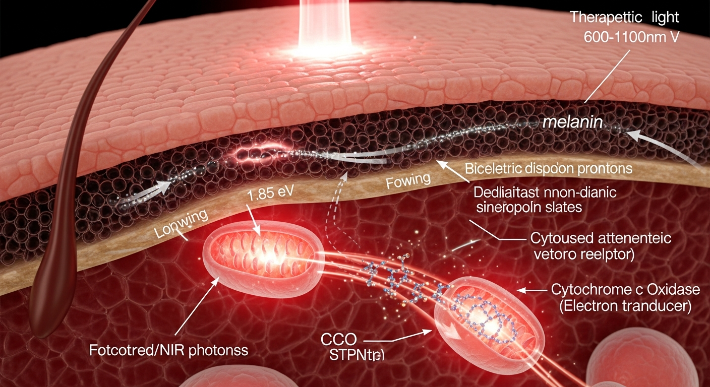
To understand the intersection of melanin and photomedicine, we must first look at the physics of light penetration in human tissue. Photobiomodulation relies heavily on the optical tissue window—a spectrum spanning approximately 600 nm to 1100 nm (red to near-infrared). Within this specific wavelength range, the absorption of light by water and hemoglobin drops significantly, allowing photons to penetrate several millimeters to centimeters into tissue.
However, melanin does not grant these photons a free pass. Unlike photosynthetic pigments like chlorophyll, which have sharp, distinct absorption peaks, eumelanin exhibits a broadband, monotonic absorption profile. As described by bio-physicist Paul Meredith and others, melanin's absorption increases exponentially toward the ultraviolet but maintains a highly significant, trailing cross-section well into the near-infrared.
When a therapeutic 810 nm laser is applied to skin classified as Fitzpatrick Type IV, V, or VI, a substantial fraction of the incident photons is immediately captured by the epidermal melanin layer. This creates a biophysical competition for photons between the superficial melanin biopolymers and the underlying target tissues. The heavily pigmented epidermis acts not just as a barrier, but as a dense optical sink, fundamentally altering the total energy that ultimately reaches deeper dermal and subdermal layers.
The Classical Target: Cytochrome c Oxidase
To appreciate why this optical competition matters, we must examine the established mechanisms of photobiomodulation. The foundational work of researchers like Tiina Karu and Michael Hamblin has demonstrated that the primary photoacceptor for red and NIR light in mammalian cells is cytochrome c oxidase (CCO), specifically Complex IV in the mitochondrial electron transport chain.
CCO contains binuclear copper centers (CuA and CuB) that absorb light efficiently in the red and NIR spectrums. When these centers absorb photons, it drives the photodissociation of inhibitory nitric oxide (NO) from the enzyme. This displacement allows oxygen to bind once again, rapidly accelerating the electron transport chain. The result is a cascade of cellular events: an increase in the mitochondrial membrane potential ($\Delta\Psi_m$), a surge in adenosine triphosphate (ATP) synthesis, and the generation of mild, transient reactive oxygen species (ROS) that activate vital transcription factors like NF-kB.
Because PBM relies on this specific photochemistry, it is governed by a strict biphasic dose-response (often modeled on the Arndt-Schulz curve). An optimal dose of photons triggers a stimulatory healing cascade, while an excessive dose causes oxidative stress and inhibits tissue function. Herein lies the melanin paradox for PBM: a light dose calibrated to optimally stimulate CCO in lightly pigmented skin may fall severely short in melanin-rich skin due to epidermal absorption. Conversely, simply turning up the power to force photons through the melanin layer risks superficial thermal damage.
Beyond the Sunscreen Model: Melanin as an Optoelectronic Transducer
If melanin is capturing a large percentage of therapeutic red and NIR photons, what exactly is it doing with that energy? Traditional dermatology views this purely as a photoprotective "sunscreen" mechanism, but biophysics tells a more complex story.
Since John McGinness's 1974 demonstration of voltage-controlled switching in melanins, we have understood that eumelanin behaves as an amorphous organic semiconductor. It possesses an estimated energy bandgap of ~1.85 eV. Notably, an energy of 1.85 eV corresponds to a photon wavelength of approximately 670 nm—falling squarely within the therapeutic window used in photobiomodulation.
When melanin absorbs these photons, greater than 99.9% of the excited state energy undergoes non-radiative relaxation through ultrafast internal conversion, safely dissipating the photon's energy as localized heat. While this protects the cell from ionizing damage, this localized photothermal event is not biologically inert. Mild, localized heating can alter cell membrane fluidity, temporarily shift cellular resting potentials, and activate temperature-sensitive transient receptor potential (TRP) ion channels. This suggests that melanin's absorption of NIR light may trigger secondary bioelectric signaling pathways in melanocytes and surrounding keratinocytes.
Furthermore, emerging theoretical frameworks propose that melanin's interaction with light extends to photochemical energy transduction. The hypotheses advanced by researchers like Arturo Solís Herrera suggest that melanin possesses the capacity to use absorbed light to drive the dissociation of cellular water, generating localized electrochemical gradients. While the extent of this water-splitting capacity remains a subject of ongoing biophysical validation, landmark studies by Albertus Mostert have definitively proven that melanin exhibits hydration-dependent proton conductivity. If red and NIR light can modulate melanin's proton-conducting state or its stable free radical populations, melanin may be actively translating phototherapy into a tangible bioelectric current at the tissue surface.
Navigating Dosimetry and the Future of Precision Photomedicine
The intersection of PBM and melanin biophysics is not merely an academic curiosity; it represents a pressing clinical challenge. Currently, the vast majority of photobiomodulation protocols and dosimetric models are established using in vitro models lacking melanin or in vivo models optimized for lighter skin phenotypes.
When translating these therapies to diverse populations, the failure to account for melanin's optoelectronic properties leads to inequitable clinical outcomes. Applying high-fluence NIR lasers to heavily pigmented skin without dosimetric adjustment can result in the rapid accumulation of heat due to melanin's highly efficient internal conversion. This not only confounds the desired biological effect but risks epidermal blistering and post-inflammatory hyperpigmentation.
To advance the field, photomedicine must evolve toward precise, individualized dosimetry. Future technologies must integrate real-time optical feedback—utilizing tools like spatial frequency domain imaging (SFDI) or optical coherence tomography (OCT)—to quantify local melanin concentrations before delivering therapy. By precisely mapping the melanin barrier, practitioners can adjust parameters such as pulse duration, irradiance, and wavelength to ensure therapeutic photon density at the mitochondrial level, while safely managing the thermal and bioelectric energy transduced by epidermal melanin.
Ultimately, we must stop viewing melanin as a biological nuisance in the path of a laser beam. It is a highly evolved, quantum-mechanically complex macromolecule. By deeply understanding how melanin absorbs, dissipates, and transduces red and near-infrared light, we open the door to novel phototherapies that work synergistically with pigmented tissues, rather than fighting against them.
Key Takeaways
- Melanin lacks a singular absorption peak, instead exhibiting a broadband absorption spectrum that heavily overlaps with the 600–1100 nm optical window used in photobiomodulation.
- The primary mechanism of PBM involves the absorption of red/NIR photons by cytochrome c oxidase, which accelerates mitochondrial respiration and ATP production, a process governed by a strict biphasic dose-response.
- As an amorphous organic semiconductor with a bandgap of ~1.85 eV, melanin's absorption of 670 nm light triggers ultrafast non-radiative relaxation, which may stimulate secondary bioelectric pathways via localized thermal gradients and ion channel activation.
- Epidermal melanin acts as a highly efficient optical sink, meaning PBM protocols calibrated for lighter skin types can result in significant under-dosing at target tissues in heavily pigmented skin.
- Advancing equitable and effective photomedicine requires integrating melanin's specific optoelectronic and photothermal properties into dynamic, personalized dosimetric models.
References
- Karu, T. I. "Primary and secondary mechanisms of action of visible to near-IR radiation on cells." Journal of Photochemistry and Photobiology B: Biology 49(1), 1-17 (1999). DOI: 10.1016/S1011-1344(98)00131-X
- Hamblin, M. R. "Mechanisms and applications of the anti-inflammatory effects of photobiomodulation." AIMS Biophysics 4(3), 337-361 (2017). DOI: 10.3934/biophy.2017.3.337
- Meredith, P., & Sarna, T. "The physical and chemical properties of eumelanin." Pigment Cell Research 19(6), 572-594 (2006). DOI: 10.1111/j.1600-0749.2006.00345.x
- McGinness, J., Corry, P., & Proctor, P. "Amorphous semiconductor switching in melanins." Science 183(4127), 853-855 (1974). DOI: 10.1126/science.183.4127.853
- Mostert, A. B., Powell, B. J., Pratt, F. L., Hanson, G. R., Sarna, T., Gentle, I. R., & Meredith, P. "Role of semiconductivity and ion transport in the electrical conduction of melanin." Proceedings of the National Academy of Sciences 109(23), 8943-8947 (2012). DOI: 10.1073/pnas.1119948109
- Barolet, D., Roberge, C. J., Auger, F. A., Boucher, A., & Germain, L. "Regulation of skin collagen metabolism in vitro using a pulsed 660 nm LED light source: clinical correlation with a single-blinded study." Journal of Investigative Dermatology 129(12), 2751-2759 (2009). DOI: 10.1038/jid.2009.186