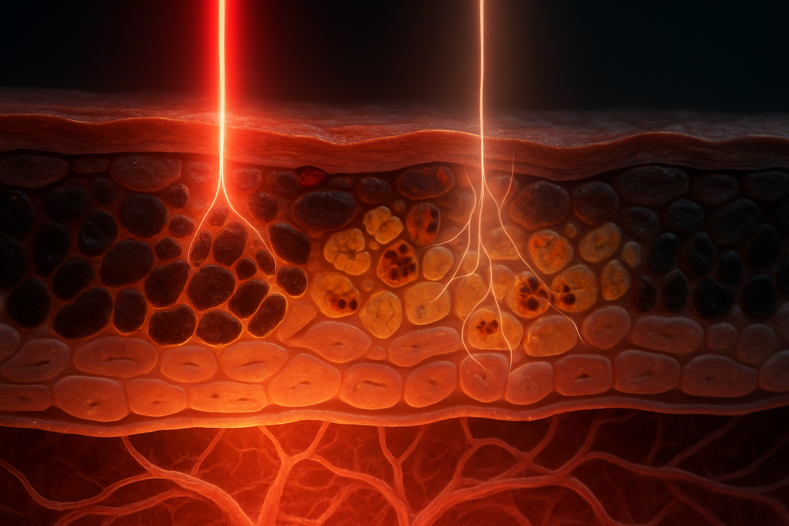
Introduction: The Role of Melanin in Photobiomodulation
Photobiomodulation (PBM) uses nonionizing red and near-infrared (NIR) light—commonly between approximately 600 and 1,100 nm—to influence cellular behavior without deliberately ablating tissue or producing substantial bulk heating. Depending on wavelength, irradiance, fluence, treatment geometry, and tissue composition, PBM has been investigated for effects on mitochondrial respiration, inflammation, pain, wound repair, nerve function, and tissue remodeling.
The central optical challenge is that the light dose delivered by a device is not necessarily the dose received by the relevant cells. A panel, laser, or light-emitting diode may deliver a precisely measured fluence at the skin surface, yet photons must still pass through the stratum corneum, epidermis, dermis, blood-containing tissue, extracellular matrix, and sometimes subcutaneous fat before reaching a proposed target. Melanin is one of the most important variable absorbers in this path, especially in the epidermis.
Melanin is not a single chemically uniform substance. Human pigmentation is determined by the amount, type, size, maturation, and intracellular distribution of melanin-containing organelles called melanosomes, as well as by how those melanosomes are transferred to and retained within keratinocytes. Eumelanin, which is brown to black, generally provides stronger broad-spectrum photoprotection. Pheomelanin, which is yellow to reddish, has different redox chemistry and can participate in oxidative processes under some conditions. The relative contribution of these pigments, their packaging within melanosomes, and their distribution across epidermal layers can influence how light is absorbed and scattered.
The interaction between PBM and melanin therefore involves at least three related processes:
- Spectral filtering: melanin absorbs a fraction of incident photons before they reach deeper cells.
- Photon transport: scattering and absorption determine how much light reaches a particular depth and how spatially uniform that light is.
- Energy and signal conversion: absorbed photons may be converted predominantly into heat, but local chemical, redox, and cellular signaling effects may also be relevant.
The practical consequence is that two individuals receiving the same surface fluence may experience different intratissue photon distributions. However, pigmentation does not automatically determine whether PBM will work or fail. A shallow epidermal target may receive considerable energy even in heavily pigmented skin, whereas a deeper target may experience a substantial reduction in fluence. The biological endpoint also matters: stimulating keratinocyte migration, reducing superficial inflammation, modulating dermal fibroblasts, and influencing a peripheral nerve are not optically equivalent objectives.
Understanding melanin's role is thus important for both experimental interpretation and treatment design. The goal is not simply to label darker skin as “less penetrable,” but to determine which wavelengths, doses, and delivery conditions produce an adequate and biologically appropriate dose at the intended target.
Melanin, Tissue Optics, and the Journey of Red/NIR Photons
Melanin: The Broad-Spectrum Absorber
Eumelanin and pheomelanin have broad, relatively featureless absorption spectra extending from ultraviolet wavelengths through the visible region and into the NIR. This broad absorption differs from the narrow spectral peaks characteristic of some purified dyes or chromophores. Melanin absorption generally decreases as wavelength increases, although the exact spectral slope depends on pigment composition, oxidation state, aggregation, hydration, and the surrounding biological matrix.
This wavelength trend explains why 630–670 nm light is generally absorbed more strongly by melanin than 800–850 nm light. It does not mean that NIR light is unaffected by pigmentation. Rather, for the same incident fluence, a smaller fraction may be absorbed in the superficial melanin-rich epidermis at 810 or 850 nm than at 660 nm. The difference can be therapeutically useful when the desired target lies several millimeters below the surface.
Melanosome organization is also important. In lightly pigmented skin, melanosomes are commonly smaller and more readily degraded within keratinocytes. In more heavily pigmented skin, melanosomes tend to be larger, more persistent, and more widely distributed throughout the keratinocyte cytoplasm. Two samples with a similar total melanin concentration can therefore exhibit different optical behavior if pigment packaging differs. Aggregation changes the effective absorbing volume and can alter local scattering, while the position of melanosomes relative to the nucleus, mitochondria, and cell membrane changes the microscopic distance over which absorbed energy and chemical signals can act.
The absorption behavior of melanin should also be distinguished from total tissue attenuation. Melanin may be the dominant variable absorber in the epidermis, but hemoglobin strongly influences visible-light transport in the dermis, particularly near its oxyhemoglobin and deoxyhemoglobin absorption bands. Water absorption becomes increasingly important at longer NIR wavelengths, especially beyond approximately 950 nm and more strongly in the 1,000–1,100 nm region. Consequently, the often-used 800–850 nm window represents a compromise: melanin and hemoglobin absorption are relatively lower than at many visible wavelengths, while water absorption has not yet become as dominant as it is at longer wavelengths.
From Photon Absorption to Energy Dissipation
The energy of an individual photon is determined by:
[ E=\frac{hc}{\lambda} ]
where (h) is Planck's constant, (c) is the speed of light, and (\lambda) is wavelength. Representative photon energies are:
- 660 nm: approximately 1.88 eV, or (3.01\times10^{-19}) J per photon;
- 810 nm: approximately 1.53 eV, or (2.45\times10^{-19}) J per photon;
- 850 nm: approximately 1.46 eV, or (2.34\times10^{-19}) J per photon.
A fluence of 1 J/cm² therefore corresponds to approximately (3.3\times10^{18}) photons/cm² at 660 nm, (4.1\times10^{18}) photons/cm² at 810 nm, and (4.3\times10^{18}) photons/cm² at 850 nm. A longer-wavelength treatment contains more photons per joule because each photon carries less energy.
These photons are nonionizing under ordinary PBM conditions. Their energies are far below those required to directly ionize biological molecules. That does not mean they are biologically inert. Absorption can alter the electronic or vibrational state of a chromophore, and the resulting excitation can be converted into heat, redox changes, altered ligand binding, or downstream signaling.
For synthetic eumelanin, the radiative relaxation quantum yield has been reported to be on the order of (10^{-3}). In practical terms, only roughly one in a thousand absorbed excitations may be released as fluorescence under the relevant experimental conditions; most of the energy is dissipated through ultrafast nonradiative pathways. This is consistent with melanin's well-known photoprotective function: it converts absorbed optical energy into lower-energy molecular motion and heat rather than allowing large amounts of damaging photochemistry to proceed.
The low fluorescence yield should not be interpreted as proof that all absorbed energy is biologically irrelevant. “Nonradiative” means that energy is not emitted as a photon. It may increase local molecular motion, influence hydrogen-bonding and hydration networks, alter redox equilibria, or contribute to very small temperature changes. At PBM irradiances, these changes are usually intended to remain below the range associated with overt thermal injury, but local nanoscale temperature gradients may not be identical to the average temperature measured at the skin surface.
For example, a treatment delivering 10 J/cm² at the surface does not deposit 10 J/cm² uniformly throughout the tissue. If 30% of the incident energy is absorbed in the superficial epidermis, that layer receives approximately 3 J/cm², while the remaining 7 J/cm² is scattered, transmitted, or absorbed elsewhere. If only 15% of the original fluence reaches a deeper target, that target receives approximately 1.5 J/cm² before accounting for absorption within the target itself. These values are illustrative rather than universal; actual fractions require wavelength-specific optical measurements.
Optical Penetration in Pigmented Skin
A simplified description of photon transport is:
[ F_{\text{target}}(\lambda)\approx F_{\text{incident}}(\lambda)e^{-\mu_{\text{eff}}(\lambda)L} ]
where (F_{\text{incident}}) is the surface fluence, (F_{\text{target}}) is the fluence at the target, (L) is the path length, and (\mu_{\text{eff}}) is an effective attenuation coefficient incorporating absorption and scattering.
In a more detailed tissue-optics treatment, the absorption coefficient (\mu_a), reduced scattering coefficient (\mu_s'), and anisotropy factor (g) are considered separately. A commonly used diffusion-style approximation is:
[ \mu_{\text{eff}}\approx \sqrt{3\mu_a(\mu_a+\mu_s')} ]
This approximation is not a complete description of every PBM geometry, particularly for thin layered tissues or highly directional beams, but it illustrates why both absorption and scattering matter. Melanin primarily increases absorption in the epidermis, while collagen, cellular structures, and refractive-index variations contribute substantially to scattering.
The commonly quoted statement that red light penetrates 1–3 mm and NIR light penetrates several millimeters should be treated as a broad order-of-magnitude description rather than a fixed anatomical boundary. “Penetration depth” may refer to a (1/e) attenuation depth, the depth at which a specified percentage of light remains, or the depth at which a biological response is observed. These are not interchangeable.
As a worked example, suppose an effective attenuation coefficient is 2 mm(^{-1}) at one wavelength. At a depth of 1 mm:
[ F(1\text{ mm})=F_0e^{-2}\approx0.135F_0 ]
Only about 13.5% of the fluence remains in this simplified model. If the coefficient is instead 1 mm(^{-1}), approximately 37% remains at 1 mm. At 3 mm, the corresponding remaining fractions are about 0.25% and 5%, respectively. Small changes in tissue optical properties can therefore produce large differences in delivered fluence at depth.
The epidermis is especially important because it may contain substantial melanin while being only roughly 50–150 micrometers thick in many body sites, although thickness varies considerably. A thin but strongly absorbing layer can remove a significant fraction of light before photons enter the dermis. The effect is greatest when melanin absorption is high and the treatment wavelength is short. At NIR wavelengths, epidermal transmission may improve, but scattering and dermal blood absorption still limit delivery.
Beam geometry introduces another complication. A collimated beam, a divergent LED source, and a broad panel do not create identical fluence distributions even when their surface irradiances are reported in the same units. In addition, scattering can cause photons to travel along paths longer than the nominal perpendicular tissue depth. The optical path length may therefore exceed the physical depth, increasing attenuation relative to a simple straight-line calculation.
Melanin may also make the superficial dose spatially heterogeneous. Melanosomes are not distributed as a perfectly uniform absorbing film; they occur within individual cells and may be concentrated around nuclei or in specific epidermal layers. At macroscopic scales this heterogeneity is averaged, but at cellular or subcellular scales some regions may receive more direct illumination than others. A conventional radiometer cannot resolve these microscopic variations.
Limitations of Skin Phototype as an Optical Proxy
Fitzpatrick phototype is useful for describing a person's tendency to burn or tan after ultraviolet exposure, but it is an imperfect substitute for measured optical properties. Phototype does not directly specify epidermal melanin concentration, melanosome size, eumelanin-to-pheomelanin ratio, blood volume, hydration, tissue thickness, or scattering coefficient.
Even within one individual, optical properties differ among anatomical sites. The palm, forearm, face, scalp, and lower leg have different epidermal thicknesses, appendage densities, blood supplies, and histories of sun exposure. Recent tanning, inflammation, vascular changes, and topical products can also modify reflectance and absorption.
A more informative optical characterization may include diffuse reflectance spectroscopy, integrating-sphere measurements, spatial-frequency-domain imaging, or time-resolved photon migration. These methods can estimate wavelength-specific absorption and scattering rather than relying on a visual category. For research, reporting measured or modeled (\mu_a), (\mu_s'), tissue thickness, melanin index, and hemoglobin-related variables would substantially improve reproducibility.
A practical example illustrates the limitation. Two participants may both be categorized as the same phototype, yet one may have a thicker epidermis with greater melanosome persistence while the other has more dermal blood volume. At 660 nm, the first participant may show stronger superficial attenuation; at a different wavelength, the second may show greater absorption from hemoglobin. The same phototype label cannot resolve these mechanisms.
Cellular Targets: From Mitochondrial Absorption to Cellular Signaling
The Role of Cytochrome C Oxidase
Cytochrome c oxidase (CCO, complex IV) is a major proposed photoacceptor in PBM. CCO is the terminal enzyme of the mitochondrial electron transport chain. It receives electrons from cytochrome c and transfers them to molecular oxygen while contributing to proton translocation across the inner mitochondrial membrane. The resulting proton-motive force supports ATP synthesis through ATP synthase.
The conventional PBM model proposes that red or NIR photons interact with copper- and heme-containing centers within CCO, changing the enzyme's functional state. One proposed mechanism is photodissociation or displacement of inhibitory nitric oxide from CCO under particular cellular conditions. If electron transport increases, proton pumping can increase, potentially raising mitochondrial membrane potential and ATP production.
This sequence is not guaranteed to occur in every cell or at every dose. Mitochondrial respiration is constrained by substrate availability, oxygen concentration, redox state, membrane integrity, and the existing rate of electron flow. A photon cannot force unlimited ATP production if oxygen or metabolic substrates are limiting. Likewise, a highly reduced respiratory chain may generate more electron leakage and ROS if electron transfer becomes mismatched with downstream demand.
Action spectra—comparisons of biological responses across wavelengths—have supported the involvement of CCO in some systems, particularly where responses resemble the absorption profile of mitochondrial chromophores. However, an action spectrum is not a molecular fingerprint by itself. Multiple chromophores can have overlapping spectral behavior, and tissue-level action spectra are shaped by wavelength-dependent penetration as well as molecular absorption.
The number of photons required for a biological response is also difficult to infer. At 10 J/cm² and 810 nm, the incident photon count is approximately:
[ \frac{10\ \text{J/cm}^2}{2.45\times10^{-19}\ \text{J/photon}} \approx4.1\times10^{19}\ \text{photons/cm}^2 ]
Only a fraction reaches mitochondria, and only a fraction of those photons is absorbed by candidate chromophores. Consequently, an incident fluence cannot be converted directly into a mitochondrial photon dose without a transport model and assumptions about chromophore concentration and absorption.
Nitric Oxide, Redox State, and Reactive Oxygen Species
Nitric oxide (NO) is a central candidate mediator because it can bind to mitochondrial respiratory components and influence vascular tone, inflammation, and cell signaling. PBM may alter the balance between NO and CCO, potentially changing respiration or increasing NO availability for signaling. The direction and magnitude of this effect may depend on oxygen tension, the initial mitochondrial state, and whether NO is generated enzymatically by nitric oxide synthases or released from bound reservoirs.
ROS are similarly context-dependent. A modest, transient increase in mitochondrial ROS can function as a second messenger. It may activate transcription factors such as nuclear factor erythroid 2–related factor 2, influence mitogen-activated protein kinase pathways, and modify inflammatory or repair programs. Excessive or prolonged ROS production, however, can damage lipids, proteins, DNA, and mitochondrial membranes.
This is one reason PBM responses often depend on dose rather than increasing indefinitely with fluence. A low dose may be insufficient to produce a detectable signal; an intermediate dose may generate a useful redox perturbation; and a high dose may produce excessive ROS, respiratory inhibition, or thermal stress. The relevant variable is not simply total ROS, but the location, duration, and chemical identity of the ROS signal.
Changes in mitochondrial activity can propagate through several pathways. Increased ATP availability may support cytoskeletal remodeling, ion pumping, protein synthesis, and cell migration. Altered redox signaling can affect inflammatory cytokine expression and antioxidant defenses. NO-related vascular effects can change local oxygen delivery. These secondary mechanisms may be more important for a tissue response than the initial photochemical event itself.
How Melanin Influences Mitochondrial Access
Melanin reduces the fluence available to cells beneath the melanin-rich epidermis. The affected targets may include basal keratinocytes, dermal fibroblasts, endothelial cells, immune cells, hair-follicle cells, peripheral nerve endings, and mitochondria within all of these populations. The reduction is wavelength-dependent and cannot be represented by a single universal correction factor.
Consider an illustrative 660 nm treatment delivering 8 J/cm² at the skin surface. If the epidermis and overlying tissue transmit 20% of that fluence to a dermal target, the target receives approximately 1.6 J/cm². If an 810 nm treatment delivers the same surface fluence but transmits 40%, the corresponding target dose would be approximately 3.2 J/cm². These percentages are hypothetical and should not be applied clinically without measurement, but they demonstrate how equal device settings can produce unequal target doses.
The intuitive conclusion that increased pigmentation always eliminates PBM responses is too simple for several reasons. First, the target may be superficial and located within or near the absorbing layer. Keratinocytes themselves can respond to light, and an epidermal response may influence deeper tissue through cytokines, growth factors, and barrier-related signaling. Second, scattering redistributes light laterally and vertically rather than merely removing it. Third, melanin absorbs energy locally, so it may alter the microenvironment of the cells containing melanosomes. Fourth, the optimal incident dose may be adjusted upward or the wavelength selected to compensate for attenuation, provided that heating and phototoxicity remain controlled.
Melanin-rich tissues may therefore display a shifted PBM dose-response curve rather than a complete absence of response. The surface fluence required to reach a given subdermal effect may be higher, while superficial effects may remain similar or even become more prominent. This distinction is important when comparing studies: a negative result in a deep tissue model may reflect inadequate target fluence, whereas a positive result in an epidermal model may not generalize to deeper structures.
Established Understanding Versus Unsolved Mysteries
CCO remains an important candidate, but it is unlikely to be the only relevant photoacceptor under all experimental conditions. Flavoproteins and other oxidized or reduced redox cofactors can absorb in overlapping spectral regions. Opsins and light-sensitive ion channels may contribute in particular cell types. Light-sensitive pathways involving calcium signaling, membrane proteins, and nitric oxide chemistry may operate independently of direct mitochondrial absorption.
Water-mediated mechanisms have also been proposed, although the exact molecular basis and physiological significance remain unsettled at common PBM wavelengths. Changes in membrane organization, interfacial water, or protein conformation could theoretically influence signaling without requiring a single dominant chromophore.
The challenge is to separate direct molecular absorption from indirect consequences of altered tissue optics. A cell-culture experiment with isolated monolayers may expose every cell to nearly the same fluence, whereas intact pigmented skin imposes a layered, scattering, oxygen-variable environment. Conversely, an in vivo response may be mediated by blood flow, immune signaling, or neural pathways rather than by direct mitochondrial absorption in the ultimate target cell.
Resolving these issues requires paired measurements: action spectra, intracellular redox indicators, oxygen consumption, mitochondrial membrane potential, NO measurements, ROS kinetics, temperature mapping, and wavelength-specific dosimetry. Experiments should also compare depigmented, naturally pigmented, and melanin-reconstituted models while controlling for cell type and tissue thickness.
Dosimetry, Response Patterns, and Choosing the Right Wavelength
Fluence and Biological Dose
Fluence is usually reported in joules per square centimeter:
[ H=E\times t ]
where (H) is fluence, (E) is irradiance in W/cm², and (t) is exposure time in seconds. For example, an irradiance of 50 mW/cm² delivered for 200 seconds produces:
[ 0.05\ \text{W/cm}^2\times200\ \text{s}=10\ \text{J/cm}^2 ]
This calculation accurately describes the surface exposure only if the irradiance is measured at the tissue plane and remains stable throughout treatment. It does not specify how much energy reaches a mitochondrial target.
A more biologically informative framework distinguishes at least four quantities:
- incident fluence at the skin surface;
- fluence entering and leaving each tissue layer;
- fluence at the target depth;
- absorbed energy within the target chromophore or cell population.
For a simplified three-layer model, suppose 10 J/cm² is incident. If the epidermis transmits 60%, the dermis transmits 50% of what enters it, and a subcutaneous target absorbs 20% of the remaining light, then the target-absorbed energy is:
[ 10\times0.60\times0.50\times0.20=0.60\ \text{J/cm}^2 ]
Again, this is a worked illustration, not a universal tissue value. It shows why surface fluence can overstate target-absorbed energy by an order of magnitude or more.
Pigmentation affects the first transmission term most strongly, but blood, water, scattering, and geometry affect the others. Reporting only the device's nominal fluence therefore makes it difficult to compare experiments performed on different skin types or anatomical sites.
The Biphasic PBM Response
PBM frequently displays a biphasic or hormetic response. A simplified response curve might contain three regions:
- Low dose: photon absorption is insufficient to generate a measurable downstream response.
- Intermediate dose: mitochondrial, redox, calcium, or transcriptional signaling produces a beneficial response.
- High dose: signaling becomes excessive, antioxidant systems are challenged, or thermal and metabolic stress reduce the response.
The optimal range is not fixed. It varies with wavelength, irradiance, treatment duration, cell type, oxygenation, metabolic state, and endpoint. A dose that increases ATP in one cell line may suppress proliferation in another. A fluence that improves wound closure in a superficial model may not deliver enough energy to a deep nerve or muscle target.
Melanin can shift the effective response curve in two different ways. For a deep target, attenuation may move the treatment leftward from the target's optimal range into its low-dose region. Increasing surface fluence could restore the target dose, but may simultaneously push the epidermis into a high-dose region. Alternatively, melanin absorption may concentrate energy in superficial cells and create a stronger epidermal response even while reducing the dose to deeper layers.
This layered dose-response problem means that “more light” is not always an adequate solution to poor penetration. A higher surface dose may increase superficial heating, erythema, or oxidative stress before the deep target reaches its optimal range. Wavelength selection, treatment area, pulse structure, and source geometry may be more effective than simply increasing fluence.
Choosing Between Red and NIR in Pigmented Tissues
Red light, typically 630–670 nm, is often useful for superficial targets because it can interact with cells in the epidermis and upper dermis. It may be appropriate when the desired effects involve keratinocytes, superficial wound edges, surface inflammation, or shallow vascular structures. However, melanin absorption is generally stronger in this region than in the 800–850 nm range, so the same surface fluence may produce less delivery to deeper tissue in highly pigmented skin.
NIR light, especially around 800–850 nm, commonly penetrates more effectively because absorption by melanin and many hemoglobin species is lower than at shorter visible wavelengths. This can make NIR attractive for targets in the deeper dermis, subcutaneous tissue, peripheral nerves, tendons, and muscle. NIR is not automatically superior: water absorption rises at longer wavelengths, and target-specific action spectra may favor another region.
Suppose a superficial target lies 0.5 mm below the surface and a deeper target lies 4 mm below it. A 660 nm source may provide adequate energy to the superficial target, while a substantial portion of its photons may be absorbed before reaching 4 mm. An 810 nm source may deliver a larger fraction to the deeper target, but its advantage could be reduced if the treatment is delivered through a highly scattering or blood-rich region. The appropriate choice depends on the depth and biology of the intended target, not on wavelength popularity alone.
Bandwidth also matters. A nominal “810 nm” LED may emit a broad distribution centered near 810 nm, while a laser may have a much narrower spectrum. If tissue absorption changes appreciably across that bandwidth, the effective transport and biological action may differ even when the central wavelength is the same.
Navigating Complex Dose Comparisons
Fluence alone does not fully define a PBM treatment. Important variables include:
- Irradiance: the same fluence can be delivered quickly at high irradiance or slowly at low irradiance.
- Exposure time: influences heat dissipation and the duration of signaling.
- Continuous versus pulsed delivery: changes peak irradiance, inter-pulse recovery, and possibly mitochondrial or neural responses.
- Beam geometry: affects divergence, focality, and spatial uniformity.
- Spot size and treatment area: influence diffusion, heat removal, and the number of cells exposed.
- Spectral bandwidth: determines the range of photon energies and absorption coefficients.
- Contact versus noncontact delivery: changes reflection losses, air gaps, and surface cooling.
- Tissue temperature and oxygenation: influence both mitochondrial respiration and the safety margin.
- Anatomical site: determines epidermal thickness, vascularity, melanin distribution, and target depth.
A robust experiment should measure irradiance at the tissue surface, verify the wavelength distribution, record exposure time and temperature, and describe the tissue model in enough detail to estimate transport. In pigmented models, reflectance or transmission measurements can reveal whether a nominally identical treatment delivers substantially different optical doses.
A useful comparison is to hold constant either the incident fluence or the estimated target fluence—but not to assume that both are constant. If two skin models transmit 25% and 50% of 810 nm light to a target, respectively, then a surface dose of 10 J/cm² produces estimated target fluences of 2.5 and 5 J/cm². To deliver approximately 5 J/cm² to both targets, the first model would require an incident fluence near 20 J/cm², assuming linear transmission and no dose-dependent changes. That adjustment might be reasonable experimentally, but it would also double superficial energy deposition in the more pigmented model. Measurements of temperature and superficial biomarkers would therefore be necessary.
Melanin Beyond Absorption: Controversies and Speculative Horizons
The Conventional Interpretation of Melanin
The conventional view treats melanin primarily as a broad-spectrum optical absorber and photoprotective filter. In this framework, melanin decreases the photon fluence that reaches deeper tissue and converts much of the absorbed energy into heat through rapid nonradiative relaxation. At PBM irradiances, the average temperature increase may be small, especially when exposure is brief and heat is removed by conduction and blood flow.
The absence of obvious bulk heating does not mean thermal effects are impossible. A useful thermal distinction is between average tissue temperature and local or transient temperature. A surface probe may report a negligible change while microscopic pigment-containing regions experience a different transient energy distribution. Whether such differences are large enough to influence ordinary PBM outcomes remains an empirical question.
Melanin's photoprotective role also means that its absorption is not inherently harmful. Absorption can prevent photons from reaching sensitive deeper structures and may reduce photochemical damage. The therapeutic problem is therefore one of dose allocation: the same absorption that protects deeper cellular components from excessive light can also prevent an intended PBM signal from reaching them.
New Hypothesis: Melanin's Redox Activity
Melanin is chemically complex and can participate in oxidation-reduction reactions, bind metal ions, and interact with free radicals. Eumelanin contains oxidizable aromatic and quinone-like structures, while pheomelanin incorporates sulfur-containing units and may have distinct redox behavior. The pigment can act as an electron donor or acceptor depending on its oxidation state and chemical environment.
These properties raise the possibility that melanin does more than filter photons. It could buffer certain radicals, alter the lifetime of reactive intermediates, bind catalytic metals, or influence the local redox state after light absorption. If PBM produces a small redox perturbation, melanin might dampen, prolong, or redirect that signal.
This hypothesis requires careful boundaries. The existence of redox-active melanin chemistry in biochemical experiments does not prove that it controls PBM responses in living human skin. Concentration, molecular accessibility, oxygen tension, pH, antioxidant capacity, and melanosome membranes all affect whether a chemical reaction is physiologically relevant. Melanin-associated redox behavior may also differ substantially between eumelanin and pheomelanin.
A useful experimental design would compare otherwise similar pigmented systems containing different pigment compositions, then measure both optical attenuation and redox outcomes. If two models transmit the same amount of light but produce different ROS or NO responses, that would support a chemical-modulator role beyond simple photon filtering. If the responses become identical after target fluence is normalized, optical attenuation would remain the more parsimonious explanation.
Hypothetical Scenarios and Their Implications
If melanin acts as a photobiological modulator, several consequences are possible. It might alter the local concentration or lifetime of ROS, influence NO chemistry, or modify how energy is distributed around melanosomes. It could also change the spatial relationship between absorbed light and neighboring mitochondria, nuclei, or membranes.
For example, a melanosome located near a mitochondrion could absorb photons and generate a short-range thermal or redox perturbation. A melanosome farther away might produce little direct influence on that mitochondrion even if the total cellular melanin content were identical. This creates a subcellular geometry problem that bulk melanin concentration cannot capture.
Pigment distribution could also influence signaling through keratinocytes. Keratinocytes containing melanin may respond to light and then release paracrine factors that affect fibroblasts, endothelial cells, or immune cells. In such a case, an observed dermal effect might not require direct NIR absorption by dermal mitochondria. It could arise from an epidermal relay.
Testing these possibilities requires more than measuring a final endpoint such as collagen expression or wound closure. Investigators would ideally measure the light field, temperature, mitochondrial respiration, ATP, membrane potential, ROS species, NO, cytokines, and cell-specific responses over time. Intratissue spectroscopy, oxygen-sensitive probes, and spatially resolved imaging could help determine whether the effect begins in melanin-containing cells or in deeper targets.
The Core Unresolved Dispute
The major unresolved dispute concerns whether CCO is the dominant photoacceptor in intact, living, melanin-rich tissue. Evidence from cell culture, isolated mitochondria, and action-spectrum studies supports a meaningful role for mitochondrial chromophores in some contexts. Yet whole skin introduces additional variables: melanin filtering, hemoglobin absorption, scattering, oxygen gradients, immune cells, vascular responses, extracellular matrix, and neural signaling.
A CCO-centered explanation may be correct for one wavelength and endpoint but incomplete for another. At low fluence, a small mitochondrial perturbation may initiate a redox signal. At higher fluence, superficial melanin-containing cells may become the principal responders. At still higher doses, thermal or oxidative mechanisms may dominate. The identity of the “primary” photoacceptor may therefore depend on experimental scale and treatment conditions.
Resolution will require experiments that independently manipulate pigment, target depth, and candidate chromophores. Useful approaches include genetically or chemically altering CCO function, blocking NO pathways, measuring mitochondrial respiration in real time, comparing different wavelengths at matched target fluence, and using tissue phantoms or reconstructed skin models with controlled melanin concentrations. Studies should distinguish surface fluence from absorbed fluence and should report whether light is delivered to isolated cells, thin tissue slices, ex vivo skin, or living intact tissue.
Concluding Insights
Melanin absorbs broadly across the visible and NIR spectrum, with absorption generally decreasing as wavelength increases. Eumelanin and pheomelanin differ chemically, and melanosome size, packaging, persistence, and intracellular distribution can affect optical behavior. Melanin-rich epidermis therefore reduces red and NIR transmission to deeper targets, but the magnitude of that reduction cannot be inferred reliably from skin color or Fitzpatrick phototype alone.
Red light near 630–670 nm is often more strongly attenuated by superficial melanin and hemoglobin, making it most naturally suited to shallow targets unless higher surface doses can be used without excessive superficial stress. NIR light near 800–850 nm often offers improved transmission into deeper tissue, although its advantage depends on scattering, blood content, water absorption, target depth, and the biological action spectrum.
Most absorbed melanin energy is dissipated through nonradiative pathways, with heat as the dominant macroscopic outcome. Nevertheless, melanin's chemical redox activity, metal binding, and ability to shape local optical and thermal environments leave open the possibility of additional photobiological effects. These possibilities remain hypotheses requiring direct testing rather than established explanations.
CCO remains the leading proposed mitochondrial photoacceptor, but PBM responses probably involve a network of chromophores and secondary pathways. NO, ROS, flavoproteins, opsins, ion channels, calcium signaling, vascular changes, and paracrine communication may all contribute depending on the tissue and endpoint.
The biphasic nature of PBM makes dosimetry especially important. A surface fluence can underestimate or overestimate the biologically relevant dose because melanin and other tissue components redistribute photons unevenly. A treatment that is optimal for superficial cells may be inadequate for deep cells, while increasing the surface dose to compensate may overdosed the epidermis. Future studies should report wavelength bandwidth, irradiance, fluence, pulse structure, beam geometry, tissue temperature, optical properties, target depth, and—where possible—estimated or measured intratissue fluence.
The most useful shift in the field is from asking whether pigmented skin “responds” to PBM toward asking which cells receive which photons, at what dose, and through which molecular pathway. Spectrometry and quantitative tissue optics should increasingly replace generalized skin typing as the basis for dosimetric comparisons. This approach would make PBM studies more reproducible and could support wavelength and dose strategies tailored to both pigmentation and anatomical target.
Related Research
- Melanin as an Energy Transducer: Converting Light, Heat, and Mechanical Energy at the Molecular Level
- What If Melanin Could Enable Direct Photosynthesis-Like Energy Production in Human Cells?
- Spin States and Radical Pairs in Melanin: The Quantum Memory Hypothesis
References
- Karu, T. I. “Primary and secondary mechanisms of action of visible to near-IR radiation on cells.” Journal of Photochemistry and Photobiology B: Biology, 1999. doi:10.1016/s1011-1344(98)00219-x
- Huang, Y.-Y., Chen, A. C.-H., Carroll, J. D., and Hamblin, M. R. “Biphasic dose response in low level light therapy—an update.” Dose-Response, 2009. doi:10.2203/dose-response.09-027.hamblin
- Jacques, S. L. “Optical properties of biological tissues: a review.” Physics in Medicine and Biology, 2013. doi:10.1088/0031-9155/58/11/r37
- Meredith, P., and Riesz, J. “Radiative relaxation quantum yields for synthetic eumelanin.” Photochemistry and Photobiology, 2004. doi:10.1562/0031-8655(2004)079<0211:rcrqyf>2.0.co;2
- Alaluf, S., Atkins, D., Barrett, K., Blount, M., Carter, N., and Heath, A. “Ethnic variation in melanin content and composition in photoexposed and photoprotected human skin.” Pigment Cell Research, 2002. doi:10.1034/j.1600-0749.2002.1o071.x