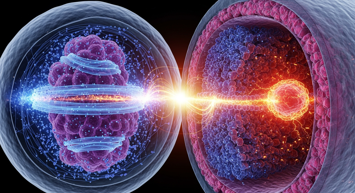
The traditional view of melanin as a passive photoprotectant is rapidly giving way to a more dynamic, biophysical paradigm. By acting as an intracellular semiconductor and proton conductor, melanin actively interfaces with the cellular membrane potential ($V_{mem}$), offering a compelling mechanism for how pigmentation regulates cell state, tissue pattern, and the threshold of oncogenesis.
For decades, the dominant textbook narrative has treated the biological cell primarily as a fluid sack of colliding biochemicals, and melanin merely as a passive anatomical parasol designed to absorb ultraviolet radiation. Yet, an expanding body of biophysical research demonstrates that biology is fundamentally bioelectric. Cells process information, regulate their cell cycles, and coordinate large-scale tissue architecture through dynamic electrical fields across their membranes. Within this electrical landscape, melanin acts not as a simple sunblock, but as a complex, electroactive polymer—a biological semiconductor capable of receiving, transducing, and transmitting electrical and ionic signals.
When we observe melanin strictly through the lens of biochemistry, we see an amorphous polymer. However, when we observe it through the lens of solid-state physics and bioelectricity, a profound intersection emerges. Emerging theoretical frameworks and established biophysical data suggest that melanin's unique capacity to conduct protons and stabilize free radicals places it in direct conversation with the electrical state of the cellular membrane. This interface—the melanin-bioelectric axis—presents a sophisticated biophysical mechanism wherein intracellular pigment actively modulates the voltage of the cell, potentially governing the delicate balance between cellular differentiation and uncontrolled proliferation.
The Bioelectric Code: Voltage as a Cellular Conductor
To understand melanin’s electrical influence, one must first examine the architecture of cellular bioelectricity. Every living cell maintains a membrane potential ($V_{mem}$)—a voltage gradient across its lipid bilayer generated by the highly regulated pumping of sodium, potassium, chloride, and protons. While neuroscientists have long studied the rapid voltage spikes in neurons, researchers have recently decoded the vital role of slow, steady electrical states in non-neural somatic cells.
Pioneering work by Michael Levin and his colleagues at Tufts University has demonstrated that $V_{mem}$ serves as a master regulator of cell behavior, guiding embryogenesis, wound healing, and tumor suppression. This bioelectric framework operates on specific voltage thresholds. Healthy, terminally differentiated cells—like mature muscle fibers or neurons—are highly hyperpolarized, typically maintaining a resting state between -60mV and -90mV. This deep negative charge acts as a bioelectric tether, locking the cell into its mature, functional identity.
Conversely, actively dividing cells, stem cells, and cancer cells are significantly depolarized. Research has identified a critical depolarization threshold at approximately -20mV. When a cell’s resting potential rises above this -20mV mark, it undergoes a profound state change. The depolarization triggers a cascade of intracellular signals that dismantle the cell's differentiated identity, plunging it back into a highly plastic, proliferative state. In Levin's Xenopus models, artificially depolarizing a tissue region is sufficient to induce oncogenesis, while hyperpolarizing established tumors can halt their growth and normalize their anatomy. The cellular voltage is not merely a byproduct of biology; it is the software that drives it.
Melanin’s Architecture: A Biological Semiconductor
How does an intracellular biopolymer interface with this bioelectric code? The answer lies in melanin’s extraordinary quantum and solid-state properties. In 1974, biophysicist John McGinness published a landmark paper in Science, demonstrating that melanin functions as an amorphous organic semiconductor. Unlike insulating biological macromolecules, melanin possesses a relatively narrow energy bandgap of ~1.85 eV, allowing for the transition of electrons between energy states upon the absorption of photons or thermal energy.
Further structural characterization by research groups led by Paul Meredith and Albertus Mostert has mapped precisely how eumelanin—the dark, photoprotective polymer formed from DHI and DHICA monomers—conducts electricity. Eumelanin is not a traditional electronic wire; rather, it is a mixed ionic-electronic conductor, behaving as a "proton glass." Its conductivity is exponentially dependent on its hydration state. Water molecules trapped within the melanin matrix dissociate, and the polymer structure facilitates rapid proton ($H^+$) hopping.
Simultaneously, melanin houses a robust population of stable free radicals, making it easily detectable via electron paramagnetic resonance (EPR) spectroscopy. This dual capacity—managing both electron clouds and proton gradients—makes melanin a highly active biophysical entity. It is an electron sink, a proton reservoir, and a dynamic capacitor capable of storing and releasing ionic charge in response to environmental stimuli.
The Melanin-Bioelectric Interface: Bridging Pigment and Potential
The theoretical convergence of melanin biophysics and membrane voltage occurs at the level of local ion gradients. The membrane potential of a cell is governed entirely by the concentration of ions inside the cell versus outside. Because melanin continuously modifies the local ionic environment through its intrinsic proton conductivity, heavily pigmented cells (such as melanocytes, retinal pigment epithelial cells, and neurons of the substantia nigra) possess a unique intracellular electrical architecture.
Consider the melanosome—the organelle that synthesizes and stores melanin—as a microscopic bio-battery. As melanin absorbs electromagnetic radiation, it generates localized proton gradients. These protons are potent modulators of cellular machinery. Fluctuations in intracellular pH directly gate specific ion channels, such as acid-sensing ion channels (ASICs) and various transient receptor potential (TRP) channels embedded in the cellular membrane. Furthermore, proton gradients dictate the state of gap junctions, the bioelectric conduits that allow neighboring cells to share voltage and coordinate their states.
By acting as an intracellular proton buffer, melanin can stabilize the cell's internal electrical environment, effectively anchoring the membrane potential. In this model, melanin functions as a bioelectric shock absorber. When a cell is subjected to oxidative stress or radiation that would typically cause ion channel dysfunction and subsequent pathological depolarization, healthy eumelanin absorbs the energetic insult, sequesters the resulting free radicals, and normalizes the local proton gradient. By doing so, melanin helps maintain the cell's hyperpolarized state (below the dangerous -20mV threshold), preserving its healthy, differentiated identity.
Pathological Depolarization: When the Axis Collapses
If healthy eumelanin stabilizes $V_{mem}$, what happens when this bioelectric axis fails? This question provides profound insights into the biophysics of melanoma and neurodegeneration.
Not all melanin is protective. Pheomelanin, the red-yellow, benzothiazine-based variant of the pigment, differs significantly in its electronic structure. It is inherently less efficient at dissipating absorbed energy and acts as a photosensitizer, actively generating reactive oxygen species (ROS) rather than quenching them. When a melanocyte accumulates excessive pheomelanin, or when its eumelanin matrix is degraded by extreme ultraviolet radiation, the polymer's capacity to regulate protons and electrons is compromised.
Under these conditions, the failing melanin polymer begins to leak oxidative stress into the cytoplasm. This oxidative burst fundamentally alters the permeability of the cellular lipid bilayer and disrupts the function of potassium and sodium pumps. The biological capacitor fails, leading to an intracellular accumulation of positive ions. The membrane potential steadily depolarizes, climbing from a healthy -60mV toward zero.
Once the melanocyte breaches the -20mV threshold, the bioelectric code dictates a state shift. The hyperpolarized signals that previously commanded the cell to remain quiescent and differentiated are silenced. Instead, voltage-sensitive morphogens and epigenetic regulators are activated, instructing the cell to proliferate aggressively and migrate. In this light, melanoma is not merely a disease of genetic mutation; it is a disease of biophysical failure. The collapse of melanin’s electronic integrity drives the collapse of the cell’s membrane voltage, ushering the cell into a cancerous state.
By recognizing the melanin-bioelectric axis, the scientific community opens a new frontier in bioelectricity and quantum biology. Understanding melanin as a localized chemical transistor that modulates cellular voltage could revolutionize our approach to pigment-related pathologies, offering new bioelectric interventions that aim to repolarize malignant melanocytes rather than simply destroying them with traditional chemotherapies.
Key Takeaways
- Cellular behavior, including differentiation and proliferation, is tightly regulated by resting membrane potential ($V_{mem}$), with a critical depolarization threshold near -20mV marking a transition toward highly proliferative, cancerous states.
- Melanin functions as an amorphous organic semiconductor with a ~1.85 eV bandgap, exhibiting mixed electronic and protonic conductivity that is highly sensitive to the polymer's hydration state.
- Through its capacity to sequester and release protons ($H^+$), melanin acts as an intracellular bio-capacitor, dynamically buffering the local pH and subsequently influencing the activity of voltage- and acid-gated ion channels.
- The presence of healthy, highly stable eumelanin likely stabilizes the hyperpolarized state of pigmented cells, acting as a bioelectric shock absorber against energetic and oxidative insults.
- The degradation of melanin or the presence of pro-oxidant pheomelanin can precipitate a bioelectric collapse, driving cellular depolarization past the -20mV threshold and potentially initiating the pathological proliferation seen in melanoma.
References
- McGinness, J., Corry, P., & Proctor, P. "Amorphous semiconductor switching in melanins." Science 183(4127), 853-855 (1974). https://doi.org/10.1126/science.183.4127.853
- Mostert, A. B., Powell, B. J., Pratt, F. L., Hanson, G. R., Sarna, T., Gentle, I. R., & Meredith, P. "Role of water in the solid-state electrical conductivity of melanin." Proceedings of the National Academy of Sciences 109(23), 8943-8947 (2012). https://doi.org/10.1073/pnas.1119948109
- Sundelacruz, S., Levin, M., & Kaplan, D. L. "Role of membrane potential in the regulation of cell proliferation and differentiation." Stem Cell Reviews and Reports 5(3), 231-246 (2009). https://doi.org/10.1007/s12015-009-9080-2
- Chernet, B. T., & Levin, M. "Transmembrane voltage potential is an essential cellular parameter for the detection and control of tumor development in a Xenopus model." Disease Models & Mechanisms 6(3), 595-607 (2013). https://doi.org/10.1242/dmm.010835
- Meredith, P., & Sarna, T. "The physical and chemical properties of eumelanin." Pigment Cell Research 19(6), 572-594 (2006). https://doi.org/10.1111/j.1600-0749.2006.00345.x
- D'Ischia, M., Wakamatsu, K., Cito, A., Panzella, L., Ito, S., & Napolitano, A. "Melanins and melanogenesis: from pigment cells to human health and technological applications." Pigment Cell & Melanoma Research 28(5), 520-544 (2015). https://doi.org/10.1111/pcmr.12393