Melanin, the ubiquitous pigment of the biological world, is most widely understood as a passive shield—a biological sunscreen that absorbs harmful ultraviolet radiation. However, a growing body of research is challenging this limited view, recasting melanin not as a simple shield, but as an active, electro-responsive biopolymer. This new perspective places melanin at the intersection of biophysics and cell signaling, suggesting it may play a direct role in modulating the fundamental electrical language of our cells.
At the heart of this emerging picture is a connection between melanin's unique semiconductor properties and the membrane potential (Vmem)—the subtle voltage difference across every cell's membrane. This is not the fast-spiking electricity of neurons, but a slower, more foundational bioelectric hum that orchestrates cell behavior, tissue development, and even disease progression. The central question now facing researchers is profound: does this ancient pigment act as a local regulator of cellular voltage, and if so, what does that mean for health, from cancer to neurodegeneration?
The Cellular Language of Voltage
Every cell in the body, not just a neuron, maintains a voltage across its outer membrane. This Vmem is established and maintained by a tightly controlled flow of ions—potassium, sodium, chloride, and calcium—through specialized protein channels. Far from being a static property, Vmem is a dynamic information-carrying medium. In his extensive work at Tufts University, Michael Levin and his colleagues have demonstrated that patterns of Vmem serve as a "bioelectric code," providing instructive cues for cells during embryonic development, regeneration, and wound healing. For instance, specific voltage gradients in a developing frog embryo pre-figure the location of the eyes, and manipulating these gradients can cause eyes to form in anatomically incorrect locations, such as the gut.
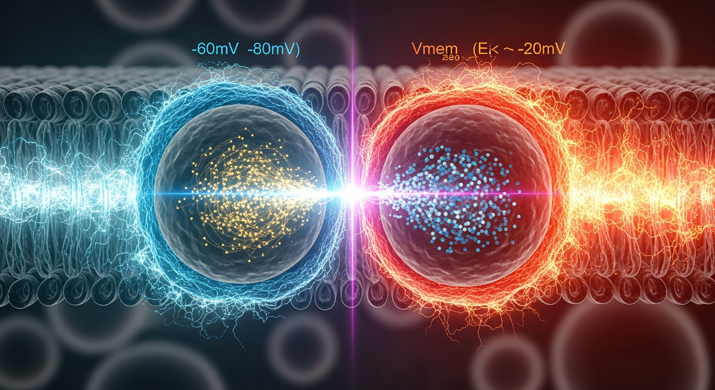
This bioelectric code also appears to play a critical role in suppressing disease. Levin's research has identified a crucial bioelectric threshold related to cell proliferation. Healthy, quiescent somatic cells typically maintain a hyperpolarized (more negative) Vmem, often between -60mV and -80mV. However, when cells undergo a sustained depolarization—a shift to a less negative voltage—their behavior changes dramatically. Specifically, Levin's group has shown that a Vmem less negative than approximately -20mV correlates strongly with a proliferative, stem-cell-like, and potentially cancerous state. This depolarization acts as a cellular trigger, unlocking transcriptional programs that lead to uncontrolled division. In this framework, cancer can be viewed, in part, as a disease of bioelectric dysregulation, where cells lose their ability to maintain a healthy, hyperpolarized state. This raises a critical question: what endogenous biological materials might help cells maintain this vital voltage?
Melanin: A Biological Semiconductor in the Cell
To understand how melanin could influence Vmem, we must look beyond its color and into its fundamental physics. Decades of research have established that eumelanin, the most common form of melanin in humans, is not an insulator but an amorphous semiconductor. In a 1974 paper in Science, John McGinness and his team first demonstrated that melanin could act as a high-resistance semiconductor capable of threshold switching, a property where its conductivity changes abruptly at a specific voltage. The accepted band gap of eumelanin—the energy required to excite an electron into a conducting state—is approximately 1.85 electron volts (eV), placing it firmly in the category of organic semiconductors.
Furthermore, melanin possesses other remarkable properties:
- Broadband Absorption: Melanin's ability to absorb energy is not limited to UV light. It absorbs across the entire electromagnetic spectrum, from UV through visible light and into the infrared, meaning it can interact with various energy sources in the cellular environment, including metabolic heat.
- Stable Free Radicals: Melanin's structure contains a population of stable free radicals, which allows it to participate in redox reactions—the transfer of electrons—without degrading. This makes it an excellent buffer for oxidative stress, but also a potential player in cellular electron transport chains.
- Proton Conductivity: Crucially, melanin is a proton conductor. Its conductivity is highly dependent on its hydration state; when hydrated, as it is within a cell, it can efficiently transport protons (H+ ions). As Vmem is directly dependent on ion gradients, including the proton gradient, this property provides a direct plausible mechanism for melanin to interface with the cell's bioelectric state.
These properties paint a picture of melanin as a sophisticated, multifunctional material. It is not a static pigment but a dynamic biopolymer capable of transducing absorbed energy (light, heat) into chemical and electrical forms, managing electron flow, and conducting protons—all processes with the potential to influence the delicate ionic balance that creates Vmem.
Connecting Pigment to Potential: The Melanin-Vmem Hypothesis
The convergence of melanin biophysics and Levin's bioelectric framework gives rise to the melanin-bioelectric axis hypothesis: the idea that intracellular melanin, particularly within organelles called melanosomes, can act as a local battery or capacitor, helping to stabilize the cell's membrane potential. While direct in vivo proof is an active area of research, several plausible mechanisms have been proposed.
One primary mechanism involves proton transport. A melanosome situated near the cell membrane could act as a "proton sponge," absorbing or releasing H+ ions. By locally altering the proton concentration, it could influence the activity of nearby proton pumps (like V-ATPases) or other ion channels in the cell membrane, thereby subtly nudging the overall Vmem towards a more hyperpolarized state. This would function as a stabilizing force, helping the cell resist the kind of pathological depolarization linked to proliferation.
A second, more speculative mechanism involves the conversion of light energy. Building on melanin's semiconductor properties, some researchers, such as Arturo Solís Herrera, have proposed that melanin can dissociate water molecules upon absorbing light, releasing electrons and protons. While this specific model of "human photosynthesis" remains debated, the underlying principle—that melanin can use light to generate charge carriers—is consistent with its semiconductor nature. This light-induced charge separation could create a localized electrical field sufficient to influence voltage-gated ion channels in the cell membrane, effectively turning the melanosome into a light-activated bioelectric regulator.
A third mechanism involves redox buffering. The electrical activity of many key ion channels is sensitive to the local redox environment (the balance of oxidizing and reducing molecules). By acting as a powerful redox buffer, melanin could stabilize the function of these channels, ensuring they operate correctly to maintain the proper hyperpolarized Vmem. In this view, melanin's antioxidant function is directly coupled to the cell's electrical health.
These proposed mechanisms suggest that melanin-rich cells may possess an additional layer of bioelectric regulation, an endogenous system for maintaining the voltage that keeps them in a quiescent, differentiated state.
From Melanoma to Morphogenesis: The Research Frontier
If the melanin-bioelectric axis is a valid biological phenomenon, the implications are significant and far-reaching.
In oncology, it offers a new lens through which to view melanoma. This aggressive skin cancer originates in melanocytes, the very cells that produce melanin. It is well-established that many melanoma cell lines exhibit a depolarized Vmem. The hypothesis suggests that the dysfunction or aberrant chemical structure of melanin in these cells might not just be a symptom of the cancer but a contributing factor to the bioelectric failure that drives it. Could therapies aimed at restoring a healthy Vmem, or modulating melanin's bioelectric function, offer a new avenue for treatment?
In neuroscience, the focus turns to neuromelanin, the form of melanin found in the substantia nigra region of the brain. The loss of these neuromelanin-containing neurons is the pathological hallmark of Parkinson's disease. While neuromelanin is known to chelate iron and protect against oxidative stress, its potential bioelectric role has been largely unexplored. These neurons have extremely high metabolic demands; perhaps neuromelanin's role is partly bioelectric, helping to stabilize the Vmem of these vulnerable cells against metabolic and oxidative insults.
Finally, in developmental and regenerative biology, the theory opens new questions. Do melanin patterns in organisms like zebrafish or salamanders play an instructive role in their development, acting as bioelectric templates alongside the Vmem gradients described by Levin? Could biomaterials engineered with melanin-like properties be used to create bioelectric scaffolds that guide tissue regeneration?
The investigation into the melanin-bioelectric axis is still in its early stages. Rigorous experiments are needed to directly measure the influence of melanosomes on the Vmem of individual cells and to untangle the precise biophysical mechanisms at play. Yet, the convergence of evidence from semiconductor physics, cell biology, and developmental bioelectricity presents a compelling case. Melanin may be far more than a simple pigment; it may be an integral component of the electrical system that patterns, powers, and protects us.
Key Takeaways
- All cells, not just neurons, use a voltage across their membrane (Vmem) as a key signaling mechanism for development, regeneration, and health.
- Research from the Levin lab at Tufts University demonstrates that a sustained Vmem depolarization to a level less negative than -20mV is a strong correlate of cell proliferation and cancerous states.
- Melanin is an organic amorphous semiconductor with properties like broadband energy absorption and hydration-dependent proton conductivity, making it a strong candidate for interacting with the cell's electrical environment.
- The melanin-bioelectric axis hypothesis proposes that melanin helps stabilize a healthy, hyperpolarized Vmem through mechanisms like local proton transport, photoelectric charge generation, or redox buffering of ion channels.
- This potential bioelectric role for melanin has profound implications for understanding melanoma, the neuroprotective function of neuromelanin in Parkinson's disease, and the bioelectric patterning of developing organisms.
- While this is an emerging field, the convergence of biophysical and bioelectric evidence suggests melanin plays an active, electro-responsive role in cell biology that is ripe for further investigation.
References
- Levin, M. "Bioelectric signaling: a primer for the unprepared." Cell & Bioscience 13(1), 32 (2023). DOI: 10.1186/s13578-023-01031-6
- McGinness, J., Corry, P., & Proctor, P. "Amorphous semiconductor switching in melanins." Science 183(4127), 853-855 (1974). DOI: 10.1126/science.183.4127.853
- Meredith, P., & Sarna, T. "The physical and chemical properties of eumelanin." Pigment Cell Research 19(6), 572-594 (2006). DOI: 10.1111/j.1600-0749.2006.00345.x
- Mostert, A. B., Rienecker, S. B., & Meredith, P. "The melanin biopolymer: a new window on solid-state electronics in the brain." Journal of Physics: Condensed Matter 29(45), 453002 (2017). DOI: 10.1088/1361-648X/aa8b56
- Chernet, B. T., & Levin, M. "Endogenous bioelectric signals as morphogenetic controls of development, regeneration, and cancer." Developmental Dynamics 242(4), 316-335 (2013). DOI: 10.1002/dvdy.23933
- Mostofian, B., et al. "Hydration-dependent proton conductivity of melanin." Proceedings of the National Academy of Sciences 113(32), 8945-8950 (2016). DOI: 10.1073/pnas.1603243113
- Zucca, F. A., et al. "Neuromelanin of the human substantia nigra: an update." Neurotoxicity Research 25(1), 13-24 (2014). DOI: 10.1007/s12640-013-9435-6
- Solís Herrera, A., Arias Esparza, M. C., et al. "The unexpected capacity of melanin to dissociate the water molecule fills the gap between the life before and after the appearance of photosynthesis." International Journal of Hydrogen Energy 35(21), 12079-12085 (2010). DOI: 10.1016/j.ijhydene.2010.08.019