Sharing an embryonic lineage with the central nervous system, melanocytes maintain vast dendritic networks throughout the epidermis. Emerging biophysical evidence suggests these cells function not merely as pigment factories, but as sophisticated bioelectric sensors mediating environmental adaptation.
Observed in isolation beneath a microscope, a melanocyte looks remarkably like a neuron. Stripped of its anatomical context, a morphologist might struggle to classify it definitively. Both cell types project long, branching dendrites into their surrounding environments. Both rely on complex vesicular transport systems to move signaling molecules along microtubule highways. Most importantly, both originate from the neural crest—a transient, pluripotent embryonic structure that migrates during development to form the peripheral nervous system, facial cartilage, and the pigment cells of the skin. Yet, while neurons are universally recognized as the body's primary computational and communication nodes, melanocytes have historically been relegated to a mechanical, singular role: synthesizing melanin to shield basal DNA from ultraviolet (UV) radiation.
This localized, purely photoprotective view is undergoing a necessary evolution. As biophysical assays become more sensitive and our understanding of endogenous cellular voltage matures, melanocytes are revealing themselves to be highly dynamic sensory sentinels. By leveraging the unique optoelectronic and chemical properties of melanin, these cells appear capable of translating environmental stimuli into complex biochemical and bioelectric actions, challenging us to rethink the functional limits of human pigmentation.
The Epidermal Melanin Unit: A Neural-Like Network
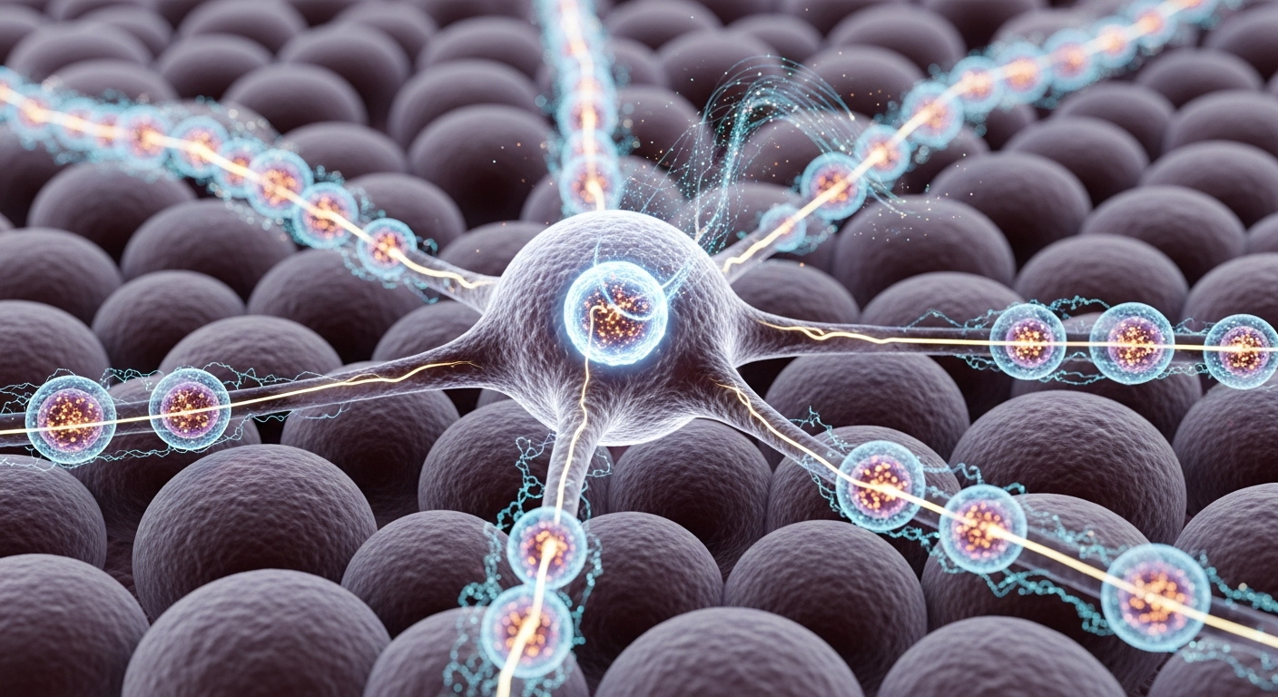
To understand the melanocyte's role beyond simple pigmentation, one must examine its structural relationship with the surrounding tissue. In human skin, melanocytes do not exist in isolation; they are the central hubs of the epidermal melanin unit. A single melanocyte extends its dendritic arms to intimately contact approximately 36 keratinocytes (the primary cell type of the epidermis).
This 1-to-36 ratio creates a highly ordered, grid-like architecture across the entire surface of the body. From a bioelectric and systems biology perspective, this morphology is virtually indistinguishable from a distributed sensor network. Research pioneered by Michael Levin and others in the field of molecular bioelectricity has established that non-neural cells utilize resting membrane potentials (Vmem) and ion channel networks to communicate positional, structural, and physiological information. Non-neural cells process information and guide morphogenesis through these electrical gradients.
Given their neural crest origin, melanocytes are uniquely positioned to participate in this bioelectric web. Their extensive dendritic arbors allow them to monitor a vast spatial territory. Rather than passively waiting for UV radiation to trigger an unregulated dump of pigment, the melanocyte continuously evaluates the physiological state of its keratinocyte network, receiving signals regarding oxidative stress, structural damage, and inflammatory cascades.
Melanosome Transfer as Intercellular Communication
The primary recognized function of the melanocyte is the production and distribution of melanosomes—specialized intracellular organelles where melanin is synthesized and stored. The orthodox view models this process as a simple delivery service: the melanocyte builds the melanosome and passes it to the keratinocyte, which positions it over the nucleus as a microscopic parasol against UV rays.
However, the mechanics of melanosome transfer suggest a much more complex form of intercellular communication. The movement of melanosomes to the dendritic tips is driven by a sophisticated motor protein complex involving Rab27a, melanophilin, and myosin Va—a biological machinery bearing striking parallels to synaptic vesicle transport in neurons.
Furthermore, melanosomes are not chemically inert packages of biological sunscreen. They possess an acidic internal pH, high concentrations of unique enzymes (like tyrosinase), stored calcium ions, and trace metals. When a melanosome is transferred via dendritic phagocytosis or direct membrane fusion, it fundamentally alters the biochemical and bioelectric state of the receiving keratinocyte. The transfer of a melanin-dense organelle represents a massive shift in local charge and redox potential. The melanosome, therefore, can be reconceptualized as a complex data packet, transmitting not just photoprotective material, but a dense payload of physiological instructions that modulates the keratinocyte's survival and differentiation pathways.
Melanin's Biophysical Signature: The Engine of the Sentinel
The key to the melanocyte's sensory capability lies in the remarkable biophysics of melanin itself. Since John McGinness and colleagues first demonstrated in 1974 that melanins function as amorphous semiconductors, researchers have labored to map the material's unique quantum and electronic behaviors.
Eumelanin, the dominant brown-black pigment in human skin, possesses a defined semiconductor bandgap of approximately 1.85 electron volts (eV). This allows the polymer to absorb broadband electromagnetic radiation across the UV and visible spectrums, efficiently dissipating 99% of absorbed photon energy as harmless heat. But melanin is also a stable free radical with an active population of unpaired electrons, rendering it highly detectable via Electron Paramagnetic Resonance (EPR) spectroscopy.
More recent work, such as that by Albertus Mostert and Paul Meredith, has highlighted melanin's capacity for proton conductivity, a property that is highly dependent on cellular hydration levels. Melanin can effectively bind, transfer, and release both electrons and protons, acting as a biological capacitor and a bidirectional redox buffer.
When situated within the living melanosome, these physical properties take on profound biological significance. Melanin is capable of interacting with reactive oxygen species (ROS), heavy metals, and electromagnetic fields. If melanin absorbs a photon or scavenges a free radical, the resulting shift in its internal charge distribution inevitably alters the local microenvironment of the organelle. It is highly probable that these biophysical changes within the melanosome cascade outward, subtly shifting the Vmem of the melanocyte. In this framework, melanin is not just a shield; it is the transducer.
The Sentinel Hypothesis: Integrating Optoelectronics and Cell Biology
By synthesizing the melanocyte's neural anatomy, its complex vesicle transport protocols, and the semiconductor physics of melanin, a new theoretical framework emerges: the Sentinel Hypothesis.
This hypothesis posits that melanocytes function as the primary environmental transducers of the integumentary system. Andrzej Slominski’s pioneering work on the neuroendocrine system of the skin has already established that the epidermis produces its own stress hormones, including Corticotropin-Releasing Hormone (CRH) and Proopiomelanocortin (POMC) derivatives. Melanocytes are active participants in this localized stress-response axis, interacting not only with keratinocytes but with local immune agents like Langerhans cells.
As environmental sentinels, melanocytes utilize their melanin reservoirs to sample the electromagnetic and chemical environment. An influx of UV radiation, a spike in localized oxidative stress, or a shift in the local electromagnetic field interacts directly with the melanin polymer. This interaction alters the protonic/electronic conductivity within the dendrites, potentially modulating the cell's ion channels and resting membrane potential. The melanocyte then transduces this biophysical data into a biological response—altering its production of neuroendocrine factors, modulating its melanosome transfer rates, and signaling the broader epidermal unit to initiate repair, inflammation, or apoptosis.
The implications for this paradigm shift are profound. By recognizing melanocytes as active, bioelectric sentinels, researchers at the Quantum Melanin Research Foundation and beyond can approach skin pathology, melanoma, and hyperpigmentation disorders not merely as defects in pigmentation, but as dysfunctions in a sophisticated biological communication network. The melanin biopolymer is the bridge between the physical universe of light and charge, and the biological realm of tissue survival.
Key Takeaways
- Melanocytes share an embryonic origin with the central nervous system (arising from the neural crest), resulting in a highly dendritic, neural-like anatomy designed for vast spatial monitoring.
- The epidermal melanin unit (one melanocyte connected to approximately 36 keratinocytes) functions as a highly organized, distributed sensor network within the skin.
- Melanosome transfer is not a passive dumping of pigment, but a highly regulated vesicular transport system—akin to synaptic transmission—that alters the receiving cell's biochemical and bioelectric state.
- Melanin's inherent biophysical properties, including its ~1.85 eV semiconductor bandgap and hydration-dependent proton conductivity, equip it to act as an environmental transducer rather than a mere physical barrier.
- The Sentinel Hypothesis models melanocytes as active bioelectric sensors that utilize melanin to detect electromagnetic and oxidative changes, translating these physical stimuli into coordinated biological stress responses.
References
McGinness, J., Corry, P., & Proctor, P. "Amorphous semiconductor switching in melanins." Science 183(4127), 853-855 (1974). DOI: 10.1126/science.183.4127.853
Mostert, A. B., Powell, B. J., Pratt, F. L., Hanson, G. R., Sarna, T., Gentle, I. R., & Meredith, P. "Role of water in the macroscopic electrical conductivity of melanin." Proceedings of the National Academy of Sciences 109(23), 8943-8947 (2012). DOI: 10.1073/pnas.1119948109
Levin, M. "Molecular bioelectricity: how endogenous voltage potentials control cell behavior and instruct pattern regulation in vivo." Molecular Biology of the Cell 25(24), 3835-3850 (2014). DOI: 10.1091/mbc.e13-12-0708
Slominski, A., Tobin, D. J., Shibahara, S., & Wortsman, J. "Melanin pigmentation in mammalian skin and its hormonal regulation." Physiological Reviews 84(4), 1155-1228 (2004). DOI: 10.1152/physrev.00044.2003
Meredith, P., & Sarna, T. "The physical and chemical properties of eumelanin." Pigment Cell Research 19(6), 572-594 (2006). DOI: 10.1111/j.1600-0749.2006.00345.x
Brenner, M., & Hearing, V. J. "The protective role of melanin against UV damage in human skin." Photochemistry and Photobiology 84(3), 539-549 (2008). DOI: 10.1111/j.1751-1097.2007.00226.x
Ando, H., Niki, Y., Ito, M., Akiyama, K., Matsui, M. S., Yarosh, D. B., & Ichihashi, M. "Melanosomes are transferred from melanocytes to keratinocytes through the processes of packaging, release, uptake, and dispersion." Journal of Investigative Dermatology 132(4), 1222-1229 (2012). DOI: 10.1038/jid.2011.413