Red and near-infrared light therapy relies on precise dosimetry to trigger mitochondrial metabolism and cellular repair. Yet, melanin's broadband absorption characteristics present a complex biophysical filter, fundamentally altering how light penetrates and interacts with pigmented tissue across diverse human phenotypes.
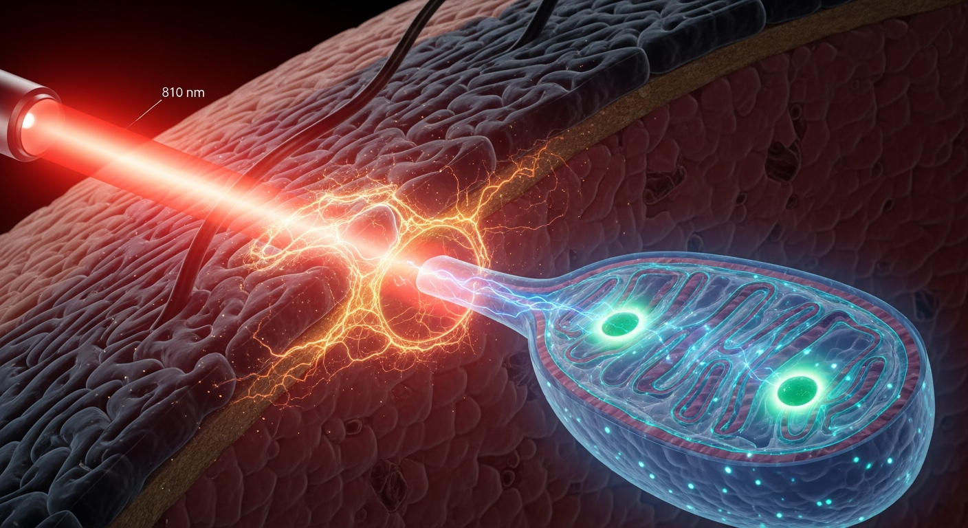
Imagine a single photon of 810-nanometer near-infrared light emitted from a clinical laser. Its intended destination lies millimeters beneath the skin's surface—perhaps the injured muscle fibers of a mammalian quad, or the compromised neurons in a superficial cortical layer. To reach its bioenergetic target, this photon must first navigate an incredibly dense, highly efficient optical filter: the epidermal melanin layer. For decades, the study of light-tissue interaction has largely treated this layer as a simple variable of attenuation, a mere barrier to be calculated and bypassed.
However, as the field of photomedicine matures, we are forced to confront a more complex biophysical reality. Melanin is not simply a biological sunshade. It is a highly conjugated biopolymer, an organic semiconductor, and an active participant in tissue thermodynamics. When we irradiate tissue with exogenous light to induce cellular healing, the melanin within the stratum basale dictates not only the volume of photons that reach deeper tissues, but also the thermal and photoacoustic landscape of the local cellular environment. Understanding this dynamic is no longer optional for rigorous photobiology—it is the central variable in translating light therapy from a generalized treatment to a precise, phenotypically tailored medicine.
The Bioenergetic Target vs. The Epidermal Filter
The foundational premise of photobiomodulation (PBM) is that specific wavelengths of red and near-infrared (NIR) light (typically between 600 and 1100 nm) can stimulate cellular functions. The established primary photoacceptor for this process is cytochrome c oxidase (CCO), known as Complex IV in the mitochondrial electron transport chain. Landmark research by photobiologist Tiina Karu demonstrated that the dinuclear copper and heme centers within CCO possess distinct absorption peaks. When these metallic centers absorb red or NIR photons, the enzyme experiences a transient conformational change. This accelerates electron transfer, boosts adenosine triphosphate (ATP) production, modulates reactive oxygen species (ROS), and initiates secondary cell-signaling cascades via calcium ion release.
Yet, to reach the mitochondria of subdermal tissues, light must first survive the epidermis. Eumelanin, the brown-black variant of melanin predominant in human skin, possesses a broad, featureless absorption spectrum. Unlike typical chromophores that absorb at specific, narrow wavelength bands, eumelanin's absorption steadily increases from the NIR down into the ultraviolet (UV) range. While its absorption coefficient is exponentially lower at 810 nm than it is at 300 nm, it is far from negligible.
In seminal optical tissue modeling by researcher Steven L. Jacques at the University of Washington, it was quantified that the absorption coefficient of a melanosome in the red/NIR therapeutic window is significant enough to severely attenuate light penetration in heavily pigmented skin. For a photon traveling toward a deep tissue target, every melanosome it encounters is an opportunity for absorption, scattering, or refraction.
The Physics of Absorption and the Biphasic Dilemma
To understand how melanin modulates PBM, we must look at what happens when a melanin molecule actually captures a red or NIR photon. Melanin is extraordinarily efficient at ultrafast internal conversion—the process of absorbing electromagnetic radiation and converting it into non-radiative phonon energy (heat) within femtoseconds. It does this with greater than 99% efficiency.
This thermodynamic reality directly complicates the core rule of PBM dosimetry: the biphasic dose-response curve, often conceptualized via the Arndt-Schulz law. PBM only works within a specific "Goldilocks" window. If the light energy (fluence) is too low, no biological stimulation occurs. If it is too high, it becomes inhibitory, potentially increasing oxidative stress and halting tissue repair.
Consider a clinical scenario treating deep tissue inflammation in a patient with a high Fitzpatrick skin type (Type V or VI), characterized by a dense concentration of melanosomes in the basal epidermis. To deliver an optimal stimulatory dose of 4 Joules per square centimeter ($J/cm^2$) to a target 2 centimeters deep, a clinician might logically increase the surface irradiance. However, the eumelanin in the epidermis will rapidly absorb this increased energy. Instead of merely passing through, the light is converted into localized heat. The intended photochemical treatment suddenly induces a photothermal effect. This not only risks epidermal damage but fundamentally alters the local cellular microenvironment, changing capillary dilation, oxygen offloading, and cellular metabolism before the light even reaches its deep-tissue destination.
Moving Beyond "One-Size-Fits-All" Dosimetry
The interaction between melanin and applied therapeutic light mandates a rigorous reassessment of standard clinical protocols. Research groups spearheaded by Michael Hamblin and others at the Wellman Center for Photomedicine have repeatedly emphasized that tissue optics must govern dosimetry.
To bypass the melanin filter, biophysicists and engineers are exploring several mechanical and optical strategies. One method involves using longer wavelengths at the absolute edge of the NIR window, such as 1064 nm Nd:YAG lasers. At this wavelength, melanin absorption drops significantly, allowing deeper penetration with reduced epidermal heating, while still engaging alternative photobiological targets (such as transient receptor potential ion channels or structured water dynamics).
Another approach relies on temporal manipulation. By utilizing super-pulsed light—delivering high peak power in ultra-short microsecond bursts followed by relatively long thermal relaxation times—researchers can "push" photons deeper into the tissue. This pulsing strategy exploits the thermal relaxation time of the melanosome. The melanin absorbs the photon but has sufficient time to dissipate the resulting heat into surrounding tissue without causing thermal damage or inhibitory signaling, allowing subsequent photons to bypass the epidermal layer safely.
Emerging Frameworks: Melanin as an Active Transducer
While the established scientific consensus correctly views melanin primarily as an optical shield that complicates the targeting of CCO, frontier research invites us to ask a bolder question: Does melanin itself play an active bioenergetic role when exposed to red and NIR light?
Melanin possesses a narrow semiconductor bandgap (approximately 1.85 eV) and exhibits stable, electron paramagnetic resonance (EPR)-detectable free radicals. Emerging theoretical frameworks, most notably those proposed by Arturo Solís Herrera regarding melanin photobiology, hypothesize that melanin does not merely dissipate light as heat, but may act as an active biological transducer. Herrera's framework suggests that melanin can use absorbed photonic energy to split water molecules, generating oxygen and molecular hydrogen—a process he controversially likens to intrinsic mammalian photosynthesis.
While Herrera’s specific hypothesis regarding water dissociation remains theoretically highly contested and outside the bounds of established consensus, the broader concept that melanin is bioelectrically active under specific light conditions is gaining biophysical traction. We know that melanin’s electrical conductivity increases exponentially with hydration, and its proton-conducting properties suggest it interacts dynamically with local electrical gradients. If melanin actively transduces absorbed NIR energy into proton or electron flow, the epidermis may not merely be a barrier to PBM—it could be a secondary, parallel target. In this emerging view, irradiating heavily pigmented skin doesn't just block light from reaching the mitochondria; it actively engages the melanin network to initiate its own unique cascade of bioelectric and thermodynamic signals.
As we advance our understanding of photomedicine, treating melanin purely as a confounding variable is scientifically insufficient. Whether acting as an extraordinarily efficient biophysical filter that dictates mitochondrial dosing, or potentially serving as an active photoacceptor in its own right, melanin is central to the efficacy of light-based therapeutics.
Key Takeaways
- Cytochrome c oxidase (CCO) is the primary established photoacceptor for red and near-infrared light, driving cellular metabolism through the electron transport chain.
- Eumelanin acts as a broadband optical filter; while its absorption peaks in the UV spectrum, it still significantly attenuates light in the 600-1100 nm therapeutic window.
- Melanin's capacity for ultrafast internal conversion transforms absorbed photons into heat, complicating photobiomodulation dosimetry by risking thermal damage at the skin surface before deep tissues receive an optimal dose.
- The biphasic dose-response of light therapy means that failing to account for melanin-induced optical attenuation will result in under-dosing deep tissues, rendering treatments ineffective for highly pigmented skin phenotypes.
- Pulsed light delivery and the use of longer near-infrared wavelengths (e.g., 1064 nm) are biophysical strategies utilized to mitigate epidermal absorption and allow for deeper photon penetration.
- Emerging theoretical frameworks suggest melanin's semiconductor and proton-conducting properties may allow it to act as an active bioelectric transducer of light, rather than existing solely as a passive optical shield.
References
- Karu TI. "Primary and secondary mechanisms of action of visible to near-IR radiation on cells." Journal of Photochemistry and Photobiology B: Biology 49(1), 1-17 (1999). DOI: 10.1016/S1011-1344(98)00128-9.
- Hamblin MR. "Mechanisms and applications of the anti-inflammatory effects of photobiomodulation." AIMS Biophysics 4(3), 337-361 (2017). DOI: 10.3934/biophy.2017.3.337.
- Jacques SL. "Optical properties of biological tissues: a review." Physics in Medicine & Biology 58(11), R37-R61 (2013). DOI: 10.1088/0031-9155/58/11/R37.
- Zein R, Selting W, Hamblin MR. "Review of light parameters and photobiomodulation efficacy: dive into complexity." Journal of Biomedical Optics 23(12), 120901 (2018). DOI: 10.1117/1.JBO.23.12.120901.
- Meredith P, Sarna T. "The physical and chemical properties of eumelanin." Pigment Cell Research 19(6), 572-594 (2006). DOI: 10.1111/j.1600-0749.2006.00345.x.
- Solís Herrera A, Arias Esparza MC, et al. "The unexpected capacity of melanin to dissociate the water molecule fills the gap between the life before and after vision." Cerebrospinal Fluid Research 7(1), S2 (2010). DOI: 10.1186/1743-8454-7-S1-S2.
- Huang YY, Chen AC, Carroll JD, Hamblin MR. "Biphasic dose response in low level light therapy." Dose-Response 7(4), 358-383 (2009). DOI: 10.2203/dose-response.09-027.Hamblin.