The Fitzpatrick scale has long been the standard for assessing UV sensitivity, but it fails to capture the rich biophysical complexity of melanin. A new approach, grounded in spectrophotometry and an understanding of melanin's semiconductor properties, could unlock a new era of personalized medicine and bioelectronic research.
In dermatology clinics and cosmetic counters worldwide, a familiar ritual unfolds. A practitioner asks a series of questions: “How does your skin respond to the sun? Do you burn easily? Do you tan?” Based on the answers, an individual is assigned a number from I to VI. This is the Fitzpatrick skin type scale, a tool so ubiquitous it has become the de facto language for describing human skin pigmentation. Developed in 1975 to determine safe starting doses for phototherapy, it serves its narrow purpose as a rough predictor of sunburn and tanning response. Yet, its very simplicity conceals a profound biophysical reality that it was never designed to address.
The scale is a proxy, a behavioral questionnaire masquerading as a biological classification. It conflates perception with physical properties and overlooks the crucial fact that two individuals with the same Fitzpatrick type can possess vastly different melanin chemistries. This discrepancy is not merely an academic detail. It carries significant implications for skin cancer risk assessment, the efficacy of light-based therapies, and our deeper understanding of melanin not just as a pigment, but as a functional, electro-active biopolymer central to cellular homeostasis. The time has come to move beyond this perceptual model and toward a quantitative, biophysical framework for classifying skin.
The Limits of a Perceptual Model
The Fitzpatrick scale was a pragmatic solution to a clinical problem. Dr. Thomas B. Fitzpatrick of Harvard Medical School needed a way to standardize dosing for PUVA (psoralen and ultraviolet A) therapy. His scale, based on self-reported sun sensitivity, provided a functional, if rudimentary, method. However, its widespread adoption has extended its use far beyond its intended scope, revealing critical limitations.
First, the scale is inherently subjective. It relies on a patient’s memory and self-assessment, which can be unreliable and culturally biased. Studies, such as one published in the Journal of the American Academy of Dermatology, have shown poor agreement between physician assessment and patient self-reporting, especially for individuals of mixed ethnic heritage who do not fit neatly into the scale's discrete categories.
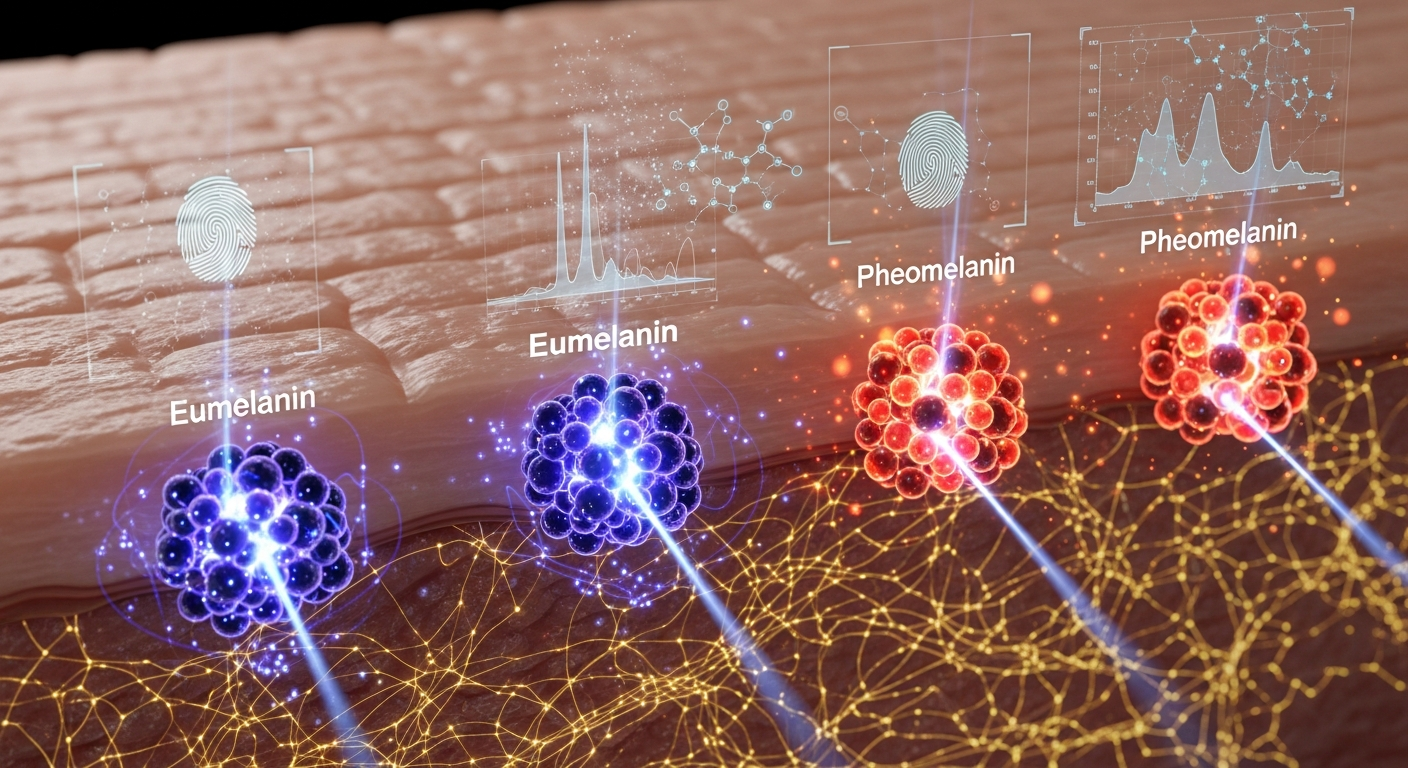
More fundamentally, the Fitzpatrick classification system is blind to the underlying chemistry of melanin. Human melanin is not a single substance but a family of polymers, primarily composed of two types: eumelanin and pheomelanin. Eumelanin, a dark brown-black polymer, is a superb photoprotectant. It absorbs a broad spectrum of UV radiation and is highly effective at dissipating that energy as heat. It is also a stable free radical, allowing it to quench reactive oxygen species (ROS). Pheomelanin, a reddish-yellow, sulfur-containing polymer, is a far less effective photoprotectant. Troublingly, upon UV absorption, pheomelanin can itself generate ROS, contributing to oxidative stress and DNA damage. The Fitzpatrick scale cannot distinguish between skin rich in protective eumelanin and skin with a high, potentially hazardous, proportion of pheomelanin.
Quantifying the Melanin Signature: A Biophysical Approach
To move beyond a subjective scale, we must directly measure the physical properties of the skin. Fortunately, the tools to do this are well-established and non-invasive. The leading technique is diffuse reflectance spectrophotometry (DRS), which illuminates a small patch of skin with light of various wavelengths and measures the light that scatters back. Because different molecules absorb light at specific wavelengths, the resulting spectrum acts as a quantitative fingerprint.
Using DRS, we can calculate a melanin index (MI), a direct measure of the total melanin content in the epidermis, far more precise than a visual assessment. More powerfully, by analyzing the shape of the absorption curve, researchers can deconvolve the relative contributions of the two melanin types. This allows for the calculation of the eumelanin-to-pheomelanin (E/P) ratio, a critical biophysical parameter that the Fitzpatrick scale completely ignores. This ratio provides a direct window into the skin's chemical and photobiological reality.
Other techniques offer even deeper insights. Electron paramagnetic resonance (EPR) spectroscopy, for instance, can directly detect and quantify the stable free radical signal inherent to eumelanin, providing a precise measure of its concentration. Raman spectroscopy can further refine the chemical signature, distinguishing the vibrational modes of different melanin subtypes. These are not futuristic technologies; they are established analytical methods in physics and chemistry that can be adapted for clinical and research settings to build a truly quantitative, multi-parametric model of skin.
From Classification to Clinical Application
Adopting a biophysical classification system is not an exercise in academic pedantry; it has immediate and profound clinical applications. The most obvious is in the field of personalized photomedicine. Current light-based therapies for conditions like psoriasis, vitiligo, and certain cancers rely on Fitzpatrick typing for dose calculation. This "one-size-fits-many" approach risks either in-effective under-dosing or painful, damaging burns. A protocol based on a measured melanin index and E/P ratio would allow clinicians to tailor the wavelength, intensity, and duration of light exposure to each patient's unique biophysical profile, maximizing therapeutic benefit while minimizing side effects.
The implications for oncology are even more significant. Decades of research by scientists like Kazumasa Wakamatsu and Shosuke Ito have established a strong link between a high pheomelanin content and increased risk for melanoma, the most dangerous form of skin cancer. The photosensitizing properties of pheomelanin contribute to a cellular environment rife with oxidative stress, a known driver of carcinogenesis. An individual with a Fitzpatrick type III, for example, might appear to have a moderate risk, but if their E/P ratio is low (i.e., high in pheomelanin), their actual risk profile may be substantially higher. Biophysical screening could identify these high-risk individuals for more intensive monitoring and preventative care long before any pathology is visible.
Looking further, a quantitative understanding of cutaneous melanin is a prerequisite for exploring its more advanced biological roles. Melanin is not an inert shield. As demonstrated by the pioneering work of John McGinness in the 1970s, eumelanin is an amorphous semiconductor with a band gap of approximately 1.85 eV, capable of conducting both electrons and protons. Its conductivity is highly dependent on its hydration state and chemical structure. This raises the possibility that melanin networks in the skin are not just for pigmentation but are part of a larger system of bioelectric signaling, potentially influencing wound healing, nerve function, and morphogenesis, echoing the principles uncovered by Michael Levin's group in developmental biology. To investigate these frontier hypotheses, we must first have the tools to accurately characterize the system's fundamental component: the melanin polymer itself. A biophysical classification system is the essential first step.
Key Takeaways
- The Fitzpatrick scale is a subjective, qualitative tool developed for predicting UV sensitivity, not for providing a comprehensive biological classification of skin.
- A biophysical approach using techniques like diffuse reflectance spectrophotometry can directly and non-invasively quantify total melanin content and, crucially, the eumelanin-to-pheomelanin (E/P) ratio.
- The E/P ratio is a far more accurate predictor of skin cancer risk and photosensitivity than visual skin type, as pheomelanin can generate damaging reactive oxygen species upon UV exposure.
- Adopting a melanin-based biophysical classification would enable personalized photomedicine, allowing for more precise and safer dosing in treatments for conditions like psoriasis and vitiligo.
- Characterizing the skin's melanin signature is a critical foundation for future research into melanin's role as a functional bio-semiconductor in bioelectric signaling, energy transduction, and tissue regeneration.
References
- Fitzpatrick, T.B. "The validity and practicality of sun-reactive skin types I through VI." Archives of Dermatology 124(6), 869-871 (1988). DOI: 10.1001/archderm.1988.01670060015008
- Eilers, S., et al. "Accuracy of self-report in assessing Fitzpatrick skin phototypes I through VI." JAMA Dermatology 149(11), 1289-1294 (2013). DOI: 10.1001/jamadermatol.2013.5353
- Kollias, N., & Baqer, A. H. "Spectroscopic characteristics of human melanin in vivo." Journal of Investigative Dermatology 85(1), 38-42 (1985). DOI: 10.1111/1523-1747.ep12276323
- Wakamatsu, K., & Ito, S. "Advanced chemical methods in melanin research." Pigment Cell Research 15(3), 174-183 (2002). DOI: 10.1034/j.1600-0749.2002.02017.x
- McGinness, J. E., Corry, P., & Proctor, P. "Amorphous semiconductor switching in melanins." Science 183(4127), 853-855 (1974). DOI: 10.1126/science.183.4127.853
- Chedekel, M. R., et al. "Photodestruction of pheomelanin: role of oxygen." Proceedings of the National Academy of Sciences 75(11), 5395-5399 (1978). DOI: 10.1073/pnas.75.11.5395
- Zecca, L., et al. "The role of iron and copper in the anabolism of neuromelanin." Journal of Neurochemistry 96(4), 1132-1138 (2006). DOI: 10.1111/j.1471-4159.2005.03541.x