Introduction: Intersecting Realms of Biology
Development transcends the mere orchestration by genetic sequences, transcription factors, or diffusible morphogens. Living tissues embody spatially organized physiological information: variations in membrane voltage, ion concentrations, gap-junctional connectivity, extracellular conductivity, mechanical tension, and cell contact. These variables can influence individual cellular behavior and, when synchronized across many cells, affect tissue-scale form.
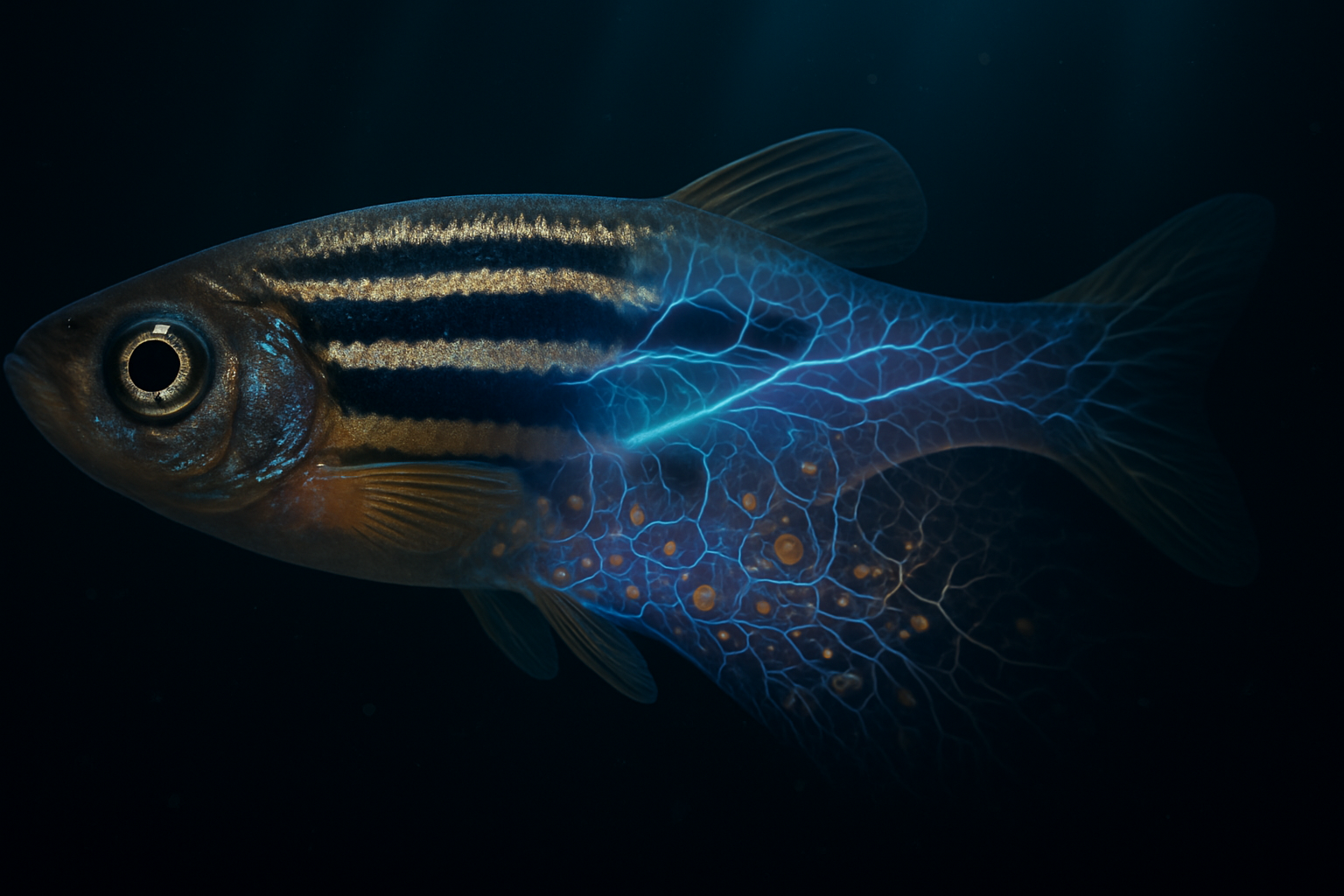
Two pertinent realms in biology have evolved in parallel. Bioelectric morphogenesis explores how membrane potentials, ion fluxes, gap junctions, and intrinsic electric fields direct processes such as migration, proliferation, differentiation, polarity, regeneration, and wound healing. Pigment-pattern morphogenesis investigates the emergence of stripes, spots, and other configurations through interactions amongst specialized pigment cells, particularly melanophores, xanthophores, and iridophores in fish skin.
The proposed "melanin template" hypothesis lies at this intersection. It questions whether patterned melanin serves as more than a visible endpoint of cellular differentiation, potentially influencing local conductivity, redox chemistry, ionic flux, or electrical potential, thus guiding tissue growth.
The distinction between tested science and hypothesis is pivotal. Bioelectric regulation of development has substantial experimental backing, and dynamic pigment-cell interactions are well-documented. Yet, the full causal sequence—melanin distribution → developmental voltage field → organ-scale growth pattern—remains elusive. Melanin might play an active role in morphogenetic electrical fields or merely mark domains established by other interactions. The Quantum Melanin Research Foundation tackles this by identifying measurable, perturbable, and testable physical links rather than considering compelling analogies as proof.
Understanding Bioelectric Signals in Morphogenesis
Membrane Potential: Informative Electricity
Cells maintain a voltage differential across their membranes, known as the resting membrane potential, (V_\mathrm{mem}). This potential, commonly ranging from −20 to −90 mV, arises from the unequal distribution of ions such as potassium, sodium, chloride, and calcium maintained by channels, pumps, and transporters like the Na(^+)/K(^+)-ATPase.
This potential isn't merely an electrical metabolic byproduct; it acts as a regulatory signal. Alterations like depolarization or hyperpolarization affect ion movement forces, channel openings, and regulate intracellular calcium. Subsequently, calcium influences processes like cytoskeletal remodeling, secretion, transcription, adhesion, and apoptosis. Voltage-dependent changes also affect pathways like Wnt, BMP, Notch, and Hippo/YAP through voltage-sensitive phosphatases modifying phosphoinositide signaling.
Voltage thus stands as a potential form of positional information. Cells can respond to electrical states, given the states' stability, spatial clarity, and coupling to molecular effectors.
Tissue-Level Electric Fields
Extracellular electric fields represent voltage gradients across tissues or extracellular spaces, emerging from ion transport, epithelial properties, damaged-cell currents, and the ionic nature of surrounding fluids or matrices. Fields around 40–200 mV/mm are significant at cellular scales, affecting migration and polarisation through mechanisms like galvanotaxis.
These fields can organize signaling complexes and adhesion machinery, translating electrical asymmetries into directed behavior.
Molecular Mechanisms: The Electric to Cellular Bridge
Several mechanisms couple electrical state to morphogenesis:
- Voltage-gated calcium channels translate depolarization into intracellular Ca(^{2+}) changes.
- Voltage-sensitive phosphatases transform membrane electric field changes into phosphoinositide metabolism alterations.
- Gap junctions facilitate ion and small signaling molecule passage, allowing tissue-level electrical coupling.
- Ion-dependent pH and osmotic variations regulate enzyme activity, cell volume, membrane trafficking, and cytoskeletal arrangement.
- Electrodiffusion links voltage gradients to movement of charged morphogens or signaling molecules.
- Transcriptional pathways involving Wnt, BMP, Notch, and Hippo/YAP respond through various intermediates like calcium and phosphorylation.
These mechanisms initiate feedback loops, where voltage changes influence cellular behavior, subsequently impacting tissue conductivity and geometry. Bioelectric morphogenesis emerges as a complex dynamical system of membranes, ions, extracellular matrices, and cellular interactions.
Evidential Boundaries
Compelling evidence exists for living cell and tissue physiology in terms of ion transport and membrane conductance. However, the presence of a developmental voltage field does not implicate melanin as the originator, nor does pigment pattern presence imply an electrical counterpart.
For a melanin-mediated mechanism to be credible, it must demonstrate that melanin or an associated cellular structure alters a relevant electrical variable, that neighboring cells detect this change, and that the resultant effect on morphogenesis is melanin-dependent, beyond contributions from pigment-cell channels or contacts.
Dynamic Pigment Patterns as Positional Architects
Chromatophore Interactions in Pigment Formation
Visible fish coloration transcends passive deposition of dark material. For instance, zebrafish adult stripe patterns involve chromatophore classes:
- Melanophores with melanin-rich melanosomes;
- Xanthophores with carotenoid pigments;
- Iridophores with guanine platelets.
These cells differ in numerous properties and interact through multicellular states rather than individual actions.
Reaction–Diffusion and Self-Patterning Phenomena
Marine angelfish studies illustrated fish pigmentation akin to a reaction–diffusion system. In such systems, local interactions can spontaneously create periodic stripes or spots.
Here, mathematical models use parameters (u) and (v) to represent interacting variables, with differential equations characterizing their dynamics. These variables might indicate chemical, cellular, electrical, or mechanical processes.
Collaborative Efforts in Pigment Evolution
In zebrafish, local xanthophore reorganization fine-tunes stripe patterns. Melanophores, xanthophores, and iridophores interact through attraction and repulsion, indicating that the pattern is a dynamic state informed by active cellular behavior.
Defining Positional Information in Pigmentation
Pigment patterns encode boundaries, spacing, and domains without inherently causing organ growth. A stripe may reflect a field without being its source. This differentiation stresses the melanin-template question, where pigment patterns correlate with voltage domains but establish no causative proof of guiding anatomical development via electrical fields.
The Melanin–Bioelectric Hypothesis: From Theory to Experimentation
Melanin as a Promising Material
Eumelanin is a disordered polymer composed of indole derivatives, showcasing broadband absorption. Depending on conditions, it is chemically reactive, redox-active, and may exhibit charge transport influenced by hydration. However, this does not make it a conventional semiconductor without clear, structurally defined band behavior.
The Hypothetical Causal Sequence
The hypothesis entails:
- Patterned melanogenesis with melanocytes producing eumelanin.
- Electrical coupling that affects local conductivity or membrane voltages.
- Voltage gradients forming stable potential differences.
- Cellular detection of these differences affecting morphogenesis.
- Integrated changes in migration, differentiation, and broader tissue adaptations.
- Organ-scale outcomes resulting in anatomical structures.
- Causal testing ensuring that disrupting melanin affects growth patterns, while restoring electrical properties rescues them.
Steps one and five are backed by pigment biology and developmental systems respectively; intermediary electrical effects remain speculative.
Consideration for Alternative Theories
Melanin may correlate with voltage without causation. Pigment cells themselves could impact fields through other biological functions. Potential segregations between melanin in melanosomes versus its spread in tissues need experimental differentiation.
Convincing Evidence Criteria
Effective experiments should control melanogenesis while tracking:
- Melanin and melanosome states;
- Membrane and extracellular voltages;
- Local ionic conditions;
- Chromatophore interactions and tissue geometry.
Causality stands if melanin removal erases voltage domains, a voltage domain restoration rescues growth, and computational models account for all interacting factors.
Pigment to Pattern: Debates and Directions
Bioelectric Signaling: Leading or Led?
Bioelectric signaling could be an organizer, a component of interactions, or an emergent consequence. It doesn't displace other patterning models but may interconnect with varied developmental contexts.
Translating Electricity to Anatomy
Voltage domains pose a translation challenge—continuous fields versus discrete anatomical details. Intermediate mechanisms could involve transcription, adhesion, migration, or calcium dynamics, amplifying initial voltage cues into sharper anatomical features.
Applying Findings Across Biological Spectra
Bioelectric regulation appears in diverse systems from amphibian to planarian models. Its ubiquity in animal chromatophore patterning demands species-specific interpretations, given differences between zebrafish skin and other organisms' tissues.
Experimental Illustration: Hypothetical Zebrafish Study
A zebrafish regeneration experiment could separate electrical contributions from pigment dynamics by altering melanogenesis and observing changes in voltage, ion conditions, and resultant tissue reconstruction through imaging and molecular mapping techniques.
The melanin hypothesis predicts electrical state changes preceding structural modifications—a confirmation that pigment patterns engage bioelectric fields in developmental biases.
Key Takeaways
- Proven Bioelectric and Pigment Morphogenesis: Both fields robustly influence cellular dynamics through established biophysical mechanisms.
- Melanin's Physical Properties Matter: With its redox-active, broadband absorbing, chemically complex nature, melanin could theoretically influence electrical fields without being a typical semiconductor.
- Unproven Complete Chain: No evidence conclusively links melanin distribution → voltage fields → growth patterns yet.
- Integrated System Exploration Needed: Methods must explore the interface of pigment-driven and bioelectric pattern influences extensively.
- The Melanin Template as Hypothesis: Currently a theorized interaction, melanin's role in bioelectric morphogenesis presents a measurable hypothesis, not an established law.
The Quantum Melanin Research Foundation seeks to bridge evidentiary gaps without overextending current theories, to unveil whether melanin-mediated charges integrate into electrodiffusive networks shaping life forms.
Related Research
- Atmospheric Melanin: Could Airborne Pigments Influence Terrestrial Bioelectrics?
- Eumelanin vs Pheomelanin: Unveiling Their Structural Differences and Biophysical Implications
- Beyond Skin Deep: A Biophysical Imperative for Melanin Classification
References
- Levin, M. (2014). “Molecular bioelectricity: how endogenous voltage potentials control cell behavior and instruct pattern regulation in vivo.” Molecular Biology of the Cell. doi:10.1091/mbc.e13-12-0708
- McCaig, C. D., Rajnicek, A. M., Song, B., & Zhao, M. (2002). “Controlling cell behavior electrically: the biophysical basis and practical implications.” Physiological Reviews, 82, 791–834.
- Kondo, S., & Asai, R. (1995). “A reaction-diffusion wave on the skin of the marine angelfish Pomacanthus.” Nature, 376, 765–768. doi:10.1038/376765a0
- Mahalwar, A., Walderich, B., Singh, A. P., & Nüsslein-Volhard, C. (2014). “Local reorganization of xanthophores fine-tunes and colors the stripe pattern of zebrafish.” Science, 345, 1242–1245. doi:10.1126/science.1254837
- Tran, M. L., Powell, B. J., & Meredith, P. (2006). “Chemical and structural disorder in the eumelanins: a possible explanation for the broadband absorbance.” Biophysical Journal, 90, 743–752. doi:10.1529/biophysj.105.069096