Deep within the human midbrain lies a region whose name translates to "black substance": the substantia nigra. Its distinctive dark hue, visible to the naked eye in post-mortem tissue, comes from a dense accumulation of a unique pigment, neuromelanin (NM). For decades, the near-total loss of this pigmented region has been recognized as the pathological hallmark of Parkinson's disease, a devastating neurodegenerative disorder. This stark correlation has long framed neuromelanin as a mere tombstone—an inert byproduct left behind by dying neurons. But a more complex and active picture is emerging from the convergence of neuroscience, biophysics, and chemistry.
The central paradox of neuromelanin is this: is it a dedicated cellular protector, a toxic catalyst for degeneration, or something far more involved in the fundamental bioenergetics of the neuron? The answer appears to be "all of the above," depending on the cell's age, its environmental stresses, and the integrity of its molecular machinery. As we look closer at this enigmatic polymer, we find it is not a passive bystander but an active participant in the life and death of the brain's most vulnerable cells, with properties that challenge our classical understanding of neuronal function.
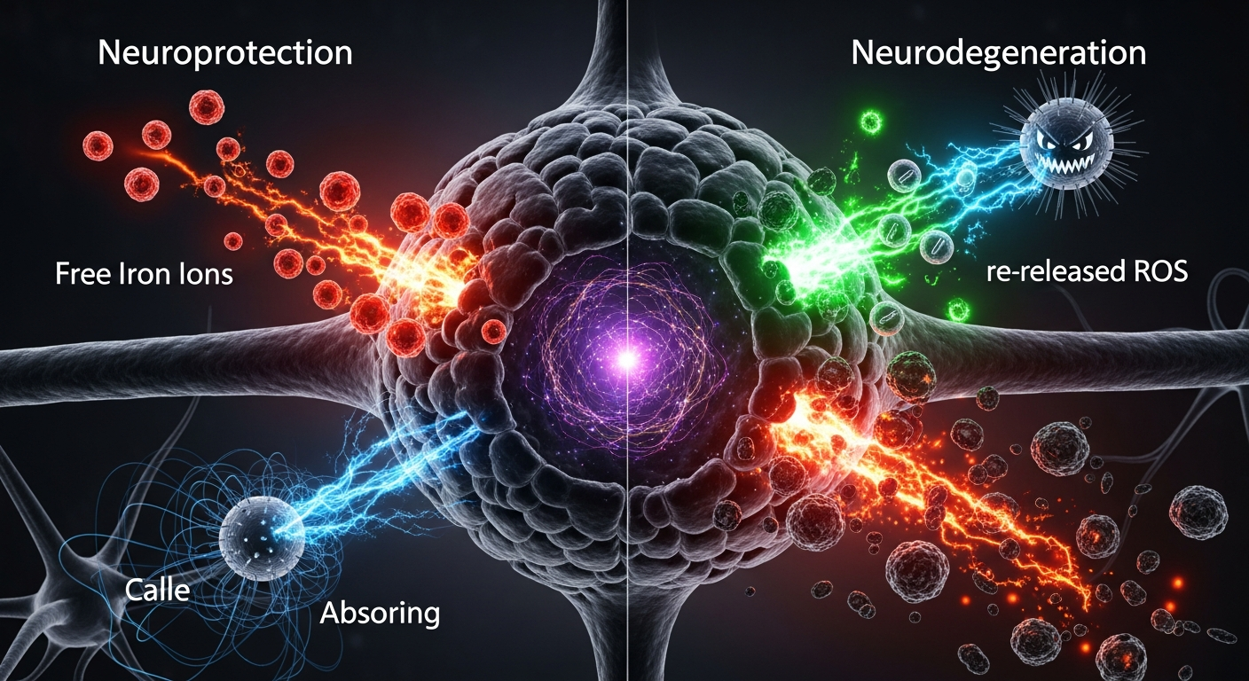
A Protective Sink in a High-Stress Environment
Neuromelanin is not synthesized in the same way as the melanin in our skin. Instead, it accumulates over a lifetime within the dopaminergic neurons of the substantia nigra pars compacta (SNc) and the noradrenergic neurons of the locus coeruleus. It is an auto-oxidative byproduct of catecholamine metabolism—specifically, the oxidation of excess cytosolic dopamine and norepinephrine. These neurons are metabolic powerhouses, firing constantly and consuming vast amounts of energy. This high metabolic rate produces a steady stream of potentially toxic byproducts, including quinones and reactive oxygen species (ROS).
In this high-stress environment, neuromelanin appears to serve as a critical cellular detoxification system. Its primary and most well-documented protective function is iron chelation. The SNc has one of the highest concentrations of iron in the brain. While essential for enzymatic function, free-form iron is highly toxic, catalyzing the production of hydroxyl radicals via the Fenton reaction—a major source of oxidative damage. Research led by pioneers like Luigi Zecca at the CNR Institute of Neuroscience in Milan has shown that neuromelanin is a remarkably high-capacity sink for iron, capable of binding it in a non-reactive state. A single NM molecule can bind hundreds of iron ions. By sequestering this dangerous metal, NM acts as a lifelong buffer, protecting the neuron from its own volatile chemistry.
Furthermore, NM can bind and sequester other toxins, including heavy metals like lead and mercury, as well as specific neurotoxins like MPP+, the active metabolite of a compound known to induce Parkinson's-like symptoms. In its healthy state, the pigment functions like a highly specialized landfill, safely containing the hazardous waste generated by a neuron's demanding existence.
The Tipping Point: When the Protector Turns Saboteur
The protective capacity of neuromelanin, however, is not infinite. The "overload hypothesis" suggests that as a neuron ages and accumulates environmental damage, its neuromelanin can become saturated with iron. Once its binding sites are full, this iron-laden neuromelanin can itself become a catalyst for ROS production, transforming the protective sink into a localized source of oxidative stress. The protector becomes a saboteur. The very molecule that defended the cell for decades may, in its final stages, accelerate its demise.
This Janus-faced role is further complicated by what happens when a neuron dies. As the cell breaks down, its neuromelanin granules are released into the extracellular space. Studies have shown that this extracellular NM is a potent inflammatory signal. It activates microglia, the brain's resident immune cells, triggering a chronic neuroinflammatory response. This microglial activation releases cytokines and other inflammatory molecules that can damage or kill neighboring, still-healthy neurons.
This process may explain the slow, progressive nature of Parkinson's disease. The death of a small number of neurons releases iron-laden NM, which triggers an inflammatory cascade that leads to the death of more neurons, creating a self-perpetuating cycle of degeneration. The physical structure of NM from Parkinson's patients is also different—smaller, more fragmented, and with a lower capacity to bind iron, suggesting a functional collapse of its protective mechanism. This transforms the pigment from a marker of cell death to an active agent in the propagation of disease.
Beyond Pathology: Bioelectric and Quantum Frontiers
While the dual role of neuromelanin in health and disease is a major area of research, its fundamental biophysical properties suggest functions that extend beyond simple chemical sequestration. Like other forms of melanin, neuromelanin is an amorphous organic semiconductor. Decades of research, dating back to the work of John McGinness and Peter Proctor in the 1970s, have established that melanins can absorb a broad spectrum of electromagnetic energy and conduct both electrons and protons.
This raises profound questions about NM's role in the living neuron. Dopaminergic neurons of the SNc are exceptionally large and have complex, unmyelinated axons, making their electrical signaling and energy management a significant challenge. Could the presence of a semiconducting polymer distributed throughout the cell body influence local electric fields or contribute to cellular charge management? The conductivity of melanin is highly dependent on its hydration state and the molecules bound to it. It is conceivable that NM's bioelectric properties are not static but are dynamically modulated by the cell's metabolic state.
Moving into more theoretical territory, neuromelanin’s unique structure invites exploration from the perspective of quantum biology. It possesses a high density of stable free radicals, which gives it a distinct paramagnetic signature detectable via electron paramagnetic resonance (EPR) spectroscopy. These stable electronic states, coupled with its ability to interact with water molecules to form ordered layers, make it a candidate for hosting coherent quantum phenomena. While highly speculative, some theoretical frameworks propose that such systems could play a role in biological information processing or energy transduction. Could the neuromelanin network within a neuron act as a form of biological memory, storing information in its electronic or structural states? Answering these questions requires new experimental approaches that can probe these properties in situ, within a living cell. This research frontier represents the core mission of the QMRF—to rigorously investigate melanin not just as a pigment, but as a functional biomaterial with extraordinary physical properties.
Key Takeaways
- Neuromelanin is a pigment that accumulates over a lifetime in dopamine- and norepinephrine-producing neurons as a byproduct of their metabolism.
- In healthy neurons, neuromelanin acts as a crucial protective agent by sequestering toxic free iron and other neurotoxins, preventing oxidative damage.
- In Parkinson's disease, it is hypothesized that neuromelanin becomes saturated with iron, transforming it from a protective molecule into a source of oxidative stress.
- When released from dying neurons, neuromelanin activates the brain's immune cells (microglia), contributing to a chronic cycle of neuroinflammation that drives disease progression.
- The loss of neuromelanin-containing neurons in the substantia nigra is a definitive pathological hallmark and diagnostic indicator for Parkinson's disease.
- Neuromelanin's established properties as an organic semiconductor and its stable free radical content suggest potential, yet unproven, roles in neuronal bioelectricity and information processing.
References
- Zecca, L., Fariello, R., Riederer, P., Sulzer, D., Gatti, A., & Tampellini, D. "The absolute concentration of neuromelanin in human substantia nigra." Brain 126(Pt 8), 1780-1786 (2003). DOI: 10.1093/brain/awg184
- Zucca, F. A., Segura-Aguilar, J., Ferrari, E., et al. "Interactions of iron, dopamine and neuromelanin in brain aging and Parkinson's disease." Progress in Neurobiology 155, 96-119 (2017). DOI: 10.1016/j.pneurobio.2015.09.012
- Zhang, W., Phillips, K., Wielgus, A. R., et al. "Neuromelanin activates microglia and induces degeneration of dopaminergic neurons: a key role for TNF-α." Neurobiology of Disease 42(3), 516-526 (2011). DOI: 10.1016/j.nbd.2011.02.012
- McGinness, J. E., Corry, P., & Proctor, P. "Amorphous semiconductor switching in melanins." Science 183(4127), 853-855 (1974). DOI: 10.1126/science.183.4127.853
- Double, K. L., Dedov, V. N., Fedorow, H., et al. "The comparative biology of neuromelanin and lipofuscin in the human brain." Cellular and Molecular Life Sciences 65(11), 1669-1682 (2008). DOI: 10.1007/s00018-008-7581-9
- Sulzer, D., Bogulavsky, J., Larsen, K. E., et al. "Neuromelanin biosynthesis in human substantia nigra." Proceedings of the National Academy of Sciences 97(22), 11852-11857 (2000). DOI: 10.1073/pnas.97.22.11852
- Fedorow, H., Tribl, F., Halliday, G., Gerlach, M., Riederer, P., & Double, K. L. "Neuromelanin in human dopamine neurons: comparison with peripheral melanins and relevance to Parkinson's disease." Progress in Neurobiology 75(2), 109-124 (2005). DOI: 10.1016/j.pneurobio.2005.02.009