The gut-brain axis is mediated not merely by biochemical neurotransmitters and microbial metabolites, but by the complex biophysics of melanin precursors. Emerging biophysical research reveals how enteric melanin, synthesized via shared pathways with central neuromelanin, offers a new lens for understanding neurodegeneration, metabolic homeostasis, and systemic bioelectricity.
When mapping the human gastrointestinal tract, biologists traditionally focus on the immense surface area of the epithelium, the diverse microbial ecologies, and the sprawling architecture of the enteric nervous system (ENS). Yet, hidden within this highly oxidative, electrically active environment is a feature that consistently evades classical gastroenterology: the physical presence of melanin and its biogenic precursors. Neural crest cells—the transient embryonic progenitors that fold and migrate to form the human nervous system—do not simply differentiate into the neurons of our brain and the melanocytes of our skin. They also migrate inward, colonizing the gut to form the 500 million neurons of the ENS, taking their melanogenic potential with them.
Why would an internal organ, devoid of ultraviolet light, maintain populations of pigment-producing cells? The answer lies in moving beyond the outdated notion of melanin as merely a biological sunscreen, and instead recognizing it as a fundamental biophysical transducer. In the gastrointestinal tract, the synthesis of melanin-like polymers from neurotransmitters, coupled with the microbiome’s tight control over precursor amino acids, suggests that melanin acts as a critical electrical and oxidative buffer. By examining the shared pathways between gut serotonin, enteric biophysics, and brain neuromelanin, we expose an integrative framework where systemic pigment biology actively modulates human health.
The Neural Crest and Enteric Melanogenesis
The architectural similarities between the brain and the gut begin at embryogenesis. Both systems rely heavily on derivatives of the neural crest. While it is well-established that these migratory cells form the enteric nervous system (ENS), histologists have also identified extracutaneous melanocytes residing in the mucosal layers of the gastrointestinal tract, the peritoneum, and the inner ear. Because these environments are entirely shielded from solar radiation, the evolutionary preservation of these cells points toward an alternative functional imperative.
Melanin, specifically eumelanin, is not an inert pigment but an organic semiconductor with a measurable bandgap of approximately ~1.85 eV. Its highly conjugated polymer structure allows it to function as an electron and proton sink. The gut is a volatile chemical reactor, subject to continuous mechanical stress, fluctuating pH, and high concentrations of reactive oxygen species (ROS) generated during digestion and immune responses. In this environment, melanin acts as an exceptional redox buffer. Because melanin maintains a population of stable free radicals—which are readily detectable via electron paramagnetic resonance (EPR) spectroscopy—it can continuously absorb and dissipate anomalous oxidative energy without being degraded. Enteric melanocytes, therefore, likely serve as localized biophysical "heat sinks," protecting the delicate mucosal and neuronal architecture of the gut from self-destruction during localized inflammatory events.
Microbiome Biosynthesis and the Precursor Economy
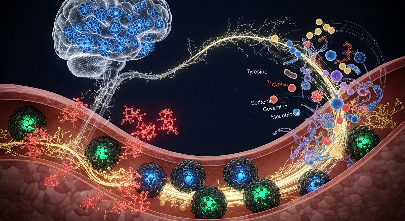
To understand the melanin-gut connection, we must examine the supply chain of its molecular precursors, a process deeply heavily heavily regulated by the gut microbiome. Both classical melanin and the specialized neuromelanin found in the central nervous system are synthesized from the oxidation of aromatic amino acids—primarily tyrosine and tryptophan—and their catecholamine derivatives, such as dopamine and serotonin.
The gut microbiome effectively acts as the central governor of this precursor economy. Groundbreaking research from institutions like Caltech, notably by researchers such as Elaine Hsiao and Jessica Yano, has demonstrated that indigenous spore-forming bacteria in the gut regulate host serotonin biosynthesis. Remarkably, up to 95% of the body’s serotonin is synthesized in the gut by enterochromaffin cells. Under conditions of oxidative stress, serotonin can undergo enzymatic or auto-oxidative polymerization to form a serotonin-melanin pathway derivative.
Microbial metabolism of tryptophan dictates whether this vital amino acid is routed toward serotonin (supporting enteric function and potential melanogenesis), the kynurenine pathway (mediating inflammation), or directly into indole derivatives. If dysbiosis shifts the metabolic processing of tyrosine and tryptophan, the systemic availability of the exact molecular building blocks required to synthesize melanin—both in the gut and eventually in the brain—is fundamentally altered. This precursor bottleneck is a critical variable in neurodegenerative disease models that are rarely analyzed through the lens of polymer biophysics.
Neuromelanin, Bioelectricity, and the Enteric Voltage
In the substantia nigra of the human brain, neuromelanin accumulates over a lifetime as a byproduct of dopamine metabolism. It plays a dual role: safely chelating transition metals like iron to prevent oxidative damage, while simultaneously posing a risk of neuroinflammation if released from dying neurons. However, the exact biophysical properties of neuromelanin-like pigments in the peripheral nervous system are an emerging frontier.
The ENS operates through complex waves of bioelectric signaling. Work spearheaded by bioelectricity pioneers like Michael Levin at Tufts University has robustly demonstrated that the membrane potential (Vmem) of non-neural and neural cells actively dictates morphogenesis, healing, and biological computation. Melanin is a unique biological material due to its hydration-dependent proton conductivity; as its hydration state changes, its ability to conduct protons shifts dramatically.
In the gut, where ion channels constantly manage extreme gradients of sodium, potassium, and chloride to drive peristalsis and secretion, the presence of melanin near enteric neurons suggests a bioelectric coupling. It is hypothesized that localized melanin deposits may act as biological capacitors, regulating the extracellular ion environment and modulating the Vmem of adjacent enteric glia and neurons. By buffering the localized bioelectric field, enteric melanin may prevent erratic depolarization in the ENS, ensuring smooth signal transduction across the gut-brain axis.
Braak’s Hypothesis and the Gut-Brain-Melanin Axis
The clinical implications of the gut-melanin axis are most evident in neurodegenerative conditions like Parkinson’s disease. In 2003, Heiko Braak and his team at Goethe University Frankfurt proposed a staging framework—now known as Braak's hypothesis—suggesting that sporadic Parkinson’s disease originates in the gut. According to this model, an unknown pathogen or stressor induces the misfolding of alpha-synuclein in the ENS, which then propagates retrograde up the vagus nerve to the brain.
Yet, Braak’s hypothesis intersects with melanin biology in ways that demand closer scrutiny. Parkinson’s disease is ultimately characterized by the profound loss of neuromelanin-pigmented dopaminergic neurons in the substantia nigra. If the disease initiates in the gut, we must ask: how does the oxidative stress in the ENS interact with enteric melanin and its catecholamine precursors before the pathology ever reaches the brain?
It is highly probable that a breakdown in the biophysical buffering capacity of the gut precedes the misfolding of proteins. If the microbiome alters the availability of tyrosine and dopamine, or if localized enteric melanocytes are depleted by severe oxidative loads, the bioelectric and redox homeostasis of the ENS collapses. The resulting oxidative storm would trigger protein misfolding, effectively making the degradation of the gut’s melanin precursor pathways the "patient zero" mechanism of catecholaminergic neurodegeneration. Understanding this axis shifts our perspective from treating neurodegeneration as an isolated brain pathology to managing it as a systemic failure of melanin biophysics and precursor metabolism.
Key Takeaways
- The neural crest gives rise to both the enteric nervous system and extracutaneous melanocytes within the gut, suggesting an evolutionary imperative for melanin in internal, UV-shielded organs.
- Eumelanin functions as an organic semiconductor and redox buffer, utilizing its stable free radicals to dissipate oxidative stress and protect the highly volatile gastrointestinal environment.
- The gut microbiome directly controls the metabolism of tyrosine and tryptophan, serving as the biological governor for the precursor molecules required to synthesize systemic and central neuromelanin.
- The synthesis of serotonin in the gut—representing 95% of the body’s supply—can feed into a serotonin-melanin pathway, producing localized polymers that sequester heavy metals and reactive oxygen species.
- Integrating melanin’s hydration-dependent proton conductivity with modern bioelectric theory suggests that enteric pigment acts as a biological capacitor, buffering the membrane potentials (Vmem) of the enteric nervous system.
- The early degradation of the gut-melanin precursor axis offers a critical, biophysically grounded expansion of Braak’s hypothesis regarding the enteric origins of Parkinson’s disease.
References
Braak, H., et al. "Staging of brain pathology related to sporadic Parkinson’s disease." Neurobiology of Aging 24(2), 197-211 (2003). DOI: 10.1016/S0197-4580(02)00065-9.
Yano, J. M., et al. "Indigenous bacteria from the gut microbiota regulate host serotonin biosynthesis." Cell 161(2), 264-276 (2015). DOI: 10.1016/j.cell.2015.02.047.
Plonka, P. M., et al. "What are melanocytes really doing all day long…? Unveiling the secret life of melanocytes." Experimental Dermatology 18(9), 799-819 (2009). DOI: 10.1111/j.1600-0625.2009.00912.x.
Double, K. L., et al. "The role of neuromelanin in neurodegeneration in Parkinson's disease." Journal of Neural Transmission 113(6), 753-766 (2006). DOI: 10.1007/s00702-006-0458-x.
Fedorow, H., et al. "Neuromelanin in human dopamine neurons: comparison with peripheral melanins and relevance to Parkinson's disease." Progress in Neurobiology 75(2), 109-124 (2005). DOI: 10.1016/j.pneurobio.2005.02.001.
Spencer, N. J., et al. "Enteric nervous system: sensory transduction, neural circuits and gastrointestinal motility." Nature Reviews Gastroenterology & Hepatology 13(4), 231-240 (2016). DOI: 10.1038/nrgastro.2016.13.
Meredith, E. J., et al. "Measurement of macroscopic and microscopic properties of melanin." Pigment Cell Research 19(6), 572-594 (2006). DOI: 10.1111/j.1600-0749.2006.00345.x.