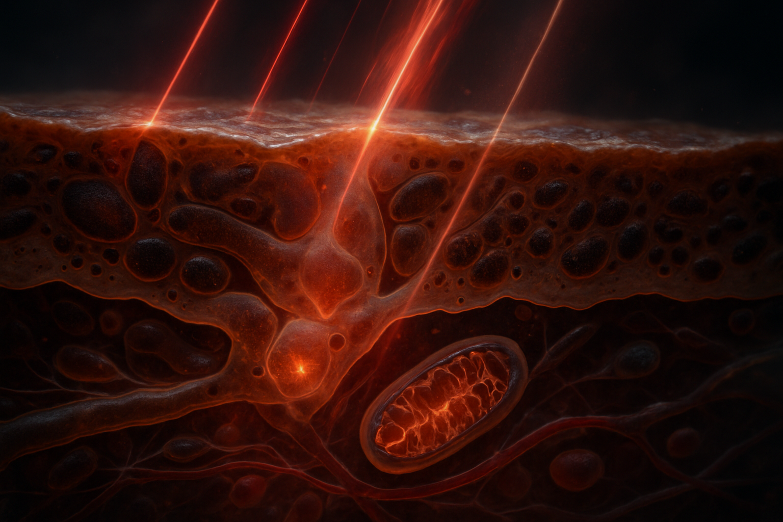
Introduction: The Significance of Pigmentation in Photobiomodulation
Photobiomodulation (PBM) employs non-ionizing red and near-infrared (NIR) light, typically within the 600–1,000 nm range, to modulate cellular functions without causing tissue damage or employing ionizing radiation. Depending on the specific parameters such as wavelength, intensity, duration, and tissue context, PBM influences a variety of biological processes including mitochondrial activity, redox balances, inflammation, vascular dynamics, neural activities, and tissue regeneration.
A primary challenge in translating PBM to clinical practice is the discrepancy between the light dose administered at the skin surface and that received by cells deeper within tissues. An emitted power of 10 J/cm² at the epidermal level may not correspond to equivalent energy exposure at deeper layers such as mitochondria within fibroblasts or vascular tissues. This discrepancy is influenced by factors including absorption, scattering, tissue geometry, blood content, and the presence of endogenous light absorbers, or chromophores.
Crucially, melanin, localized in epidermal melanosomes, serves as a broadband absorber of visible and NIR light, impacting photon penetration and subsequently altering energy distribution through absorption spectra. Depending on concentration, cellular distribution, and variant type (eumelanin or pheomelanin), melanin modulates PBM outcomes by filtering photons and converting energy into heat. The pertinent inquiry is whether melanin simply obstructs PBM, or it modulates PBM via mechanisms such as photon attenuation, spectral filtering, local thermal deposition, redox activity, and potential photochemical signaling. Despite strong evidence for its role in altering tissue optics, research continues to explore melanin’s effects on cellular mechanisms. The Quantum Melanin Research Foundation prioritizes objective assessment of pigmentation in complex biophysical studies, extending beyond its traditional classification as a superficial trait.
The Optical Pathways: Navigating Red and NIR Light through the Skin
Melanin as a Broadband Absorber
Both eumelanin and pheomelanin exhibit extensive absorption spectra from the ultraviolet through to the NIR spectrum. Unlike a distinct molecular absorption line, melanin's absorption manifests broadly, diminishing toward longer wavelengths, such that 630–660 nm light is absorbed more effectively than 810–850 nm light. This doesn’t negate NIR attenuation—skin remains a optically complex medium with contributions from hemoglobin, water, along with structural compounds such as collagen and keratin which all affect light scattering and absorption. Therefore, the internal fluence relies on radiative transfer through a multi-layered tissue, demonstrating more than just a melanin influence.
Anatomy and Melanosome Dynamics
Melanin is primarily housed within melanosomes of epidermal melanocytes and gets transferred to keratinocytes. These organelles create a non-uniform optical layer, and their placement either above or enveloping keratinocyte nuclei affects how light is absorbed. Hence, skin pigmentation is influenced by:
- Epidermal melanin levels;
- Melanosome density, size, and configuration;
- Eumelanin-to-pheomelanin ratio;
- Epidermal thickness and location;
- Water content and blood flow;
- Dermal scattering behavior.
As most melanosomes reside near the skin’s surface, they predominantly temper photon penetration before deeper layers such as fibroblasts are reached. Therefore, varying pigment levels result in disparate PBM regimes within the same tissue.
Impact of Wavelength on Energy and Penetration
Photon energy correlates with wavelength, with 660 nm photons carrying approximately 1.88 eV and 830 nm photons about 1.49 eV. Greater photon energy does not inherently translate to superior biological outcomes. Effective PBM depends on factors like absorption likelihood, cellular accessibility, light intensity, and physiological state of target cells. Typical penetration depths for skin are 1–3 mm at 630–660 nm and 2–5 mm at 800–850 nm, but these are modulated by melanin levels, tissue architecture, and other physiological factors.
From Surface to Cellular Doses
Incident fluence is measured as energy per square centimeter at the treatment surface, while target-cell fluence describes the energy that actually penetrates cells after accounting for absorption and scattering. The two can vastly differ based on tissue environment. For example, efficient dermal response in lightly pigmented skin versus epidermal energy absorption in darker skin highlights that quantitative fluence alone does not dictate biological responses.
Established PBM Biology: Identifying Photoacceptors and Signaling Pathways
Cytochrome c Oxidase (CCO): A Historical Mechanism
Cytochrome c oxidase (CCO), part of the electron transport chain, has long been seen as a key photoacceptor within the red/NIR PBM paradigm. Its overlapping absorption characteristics with red/NIR light, especially around 620–680 nm and 760–900 nm, suggest its notable role in PBM. However, absorption does not guarantee predominant influence in intact tissues, given the complex interplay between chromophores and cellular structures.
Mitochondrial Responses and Hypotheses
Red/NIR exposure is thought to modulate:
- Electron transport chains;
- Membrane potential;
- ATP productivity;
- Nitric oxide dynamics within CCO;
- ROS levels;
- Pathways sensitive to redox changes.
Initial optical perturbations lead to downstream signaling cascades influencing cellular metabolism, growth factors, inflammation, and tissue restoration. Slight ROS elevation could aid cellular signaling, but excessive levels might cause cell damage—a potential explanation for biphasic responses to PBM.
Beyond CCO: Additional Chromophore Candidates and Mechanisms
Other photoacceptors (flavoproteins, opsins, ion channels) and pathways proposed include membrane conductance changes, calcium fluxes, and reactions mediated by endogenous flavins. PBM could involve a collective response via various chromophores—these dynamics vary by cellular environment, state of health, and experimental setup.
CCO as a Plausible Component, Not the Sole Architect
Significant uncertainties complicate the CCO-centric model. Observations such as subtle CCO absorption shifts, nitric oxide dispersion conditions, indirect effects via thermal or membrane channels emphasize the need for more comprehensive mechanisms that include melanin’s role in modulating light distribution and signaling pathways at cellular levels.
Melanin’s Multifaceted Role: Optical Filter, Heat Source, or Signaling Modulator?
Photon Attenuation and Spectral Filtering
Melanin reliably performs in light attenuation by absorbing photons within epidermal layers, more so for shorter red wavelengths. Such filtering not only reduces tissue fluence but also alters the spectral energy distribution reaching deeper tissues, modulating how cells and mitochondria access these photons.
Nonradiative Energy Transfer and Heating
Melanin-mediated nonradiative relaxation leads to localized heat production, primarily through eumelanin, albeit the relevance of this process to PBM’s therapeutic effects requires further exploration. Heat may alter cellular membranes or cascade biochemical responses while mitochondrial redox reactions operate concurrently.
Redox and Chemical Properties
Chemical attributes of melanin allow it to participate in redox processes, though their exact contribution to PBM remains undetermined. While distinct forms of melanin (eumelanin and pheomelanin) possess unique oxidative behaviors, definitive evidence for direct engagement in PBM photochemical signaling is scarce.
From Passive Filtering to Active Participation?
Collected data suggest a hierarchy:
- Established knowledge: Melanin significantly alters light transmission.
- Theoretical proposals: Melanin influences local thermal conditions.
- Scarce direct insight: Melanin as a primary PBM signaling mediator remains speculative.
Melanin’s role varies by target and depth, especially when identifying changes in PBM pathways in response to pigmentation. Pigmented tissue may shift both focal energy and cellular response, demanding re-evaluation of how we perceive melanin's inhibitory potential.
Dose Variability, Biphasic Dynamics, and Pigmentation-Aware PBM Innovations
Recognized Science: Biphasic Dose Relationships in PBM
PBM's efficacy often presents a biphasic dose-response. Subthreshold doses yield minimal effects; viable doses amplify signaling, repair, and inflammatory relief; excessive doses reduce benefits, with doses typically spanning 0.1–10 J/cm² within PBM literature, underscoring the non-linearity and fine calibration necessary for optimal PBM application.
Recognized Science: Wavelength-Specific Penetration and Effects
Red and NIR wavelengths pose distinct clinical considerations. While 630–660 nm targets shallow structures, 810–850 nm provides deeper penetration with reduced melanin interference. Titling NIR as universally advantageous overlooks variations in tissue and biological responses necessitating careful wavelength tailoring per therapeutic goal.
Case Study Proposal Hypothetically
A prospective study might systematically compare PBM in variable melanin contexts using 660 nm and 830 nm beams at a 10 J/cm² dose for:
- Reflectance and transmittance analyses;
- Temperature gradients at varying tissue depths;
- Fibroblast ATP production beneath reconstructed epidermis;
- Biochemical signaling cascades.
Data may reveal stronger light attenuation in darker samples at 660 nm, altering energy localization within epidermal layers—an optical basis, rather than resistance, for observed variances in treatment efficacy.
Gaps Identified: From Surface Dose to Personalized Dose Precision
PBM methodology lacks standardization concerning specific tissue dose measurements, often omitting considerations of:
- Precise epidermal melanin analytics;
- Accurate photosynthetic absorption coefficients;
- Temperature-dependent effects.
Large-scale, objectively analyzed pigmentation trials are few. Although Fitzpatrick grading is routine cosmetically, it lacks precision in defining actual pigmentation and associated optical traits, promoting the potential for customized PBM interventions by aligning optical assessments with individual dermal profiles.
Envisioning Personalized Optical PBM Paradigms
Progressing towards personalized PBM could allow optimization beyond standard skin dose prescriptions to target-cell dosages. Adjustments could harness 810–850 nm for deeper melanin-rich tissues, refine treatment duration through empirical data, and incorporate real-time feedback for thermal management—pending sufficient confirmatory research validating its efficacy and safety.
This approach underscores melanin’s role in dictating the location and effect of PBM signals, affirming its relevance not just in physical light interactions, but as a potential orchestrator of deeper biological mechanisms.
Key Insights
Melanin’s robust light absorption modulates PBM, attenuating red wavelengths more significantly than those near 800–850 nm. This feature, together with melanosome properties, scattering, and blood characteristics, requires nuanced understanding for effective subsurface and intracellular energy management.
While providing consistent optical modulation, melanin’s heat-producing potential and signaling intricacies remain theoretical, with cytochrome c oxidase highlighted as a non-exclusive photoacceptor and PBM mediator. Given the dose-dependent biphasic response of PBM, mere fluence amplification isn’t adequate without clarity on cellular targeting and thermal effects.
Focused research should emphasize cross-group optical profiling, integrating red and NIR evaluations under objective thermal and melanin profiling. This aligns with Quantum Melanin Research Foundation’s commitment to advancing pigmentation-inclusive biophysics, though such practice must evolve beyond conjecture to an established health science framework.
Related Research
- Melanin-Based Organic Electrochemical Transistors: The Future of Biocompatible Computing
- [Melanin as a Biological Semiconductor: Bridging Quantum Physics and Cellular Biology](/articles/melanin-as-a-biological-semiconductor-bridging-quantum-physics-and-cellular-biology-1786976946135)
- What If Melanin Acts as a Biological Antenna for Environmental Electromagnetic Information?
References
- Karu, T. I. “Primary and secondary mechanisms of action of visible to near-IR radiation on cells.” Journal of Photochemistry and Photobiology B: Biology. 1999;49(1):1–17. doi:10.1016/s1011-1344(98)00219-x
- Huang, Y.-Y., Chen, A. C.-H., Carroll, J. D., and Hamblin, M. R. “Biphasic dose response in low level light therapy.” Dose-Response. 2009;7(4):358–383. doi:10.2203/dose-response.09-027.hamblin
- Jacques, S. L. “Optical properties of biological tissues: a review.” Physics in Medicine and Biology. 2013;58(11):R37–R61. doi:10.1088/0031-9155/58/11/r37
- Meredith, P., and Sarna, T. “The physical and chemical properties of eumelanin.” Pigment Cell Research. 2006;19(6):572–594. doi:10.1111/j.1600-0749.2006.00345.x
- Anderson, R. R., and Parrish, J. A. “The optics of human skin.” Journal of Investigative Dermatology. 1981;77(1):13–19. doi:10.1111/1523-1747.ep12479191