Melanin is classically understood as a photoprotective pigment, yet melanin-producing cells are distributed throughout the absolute darkness of the human gastrointestinal tract. Emerging research into the gut-brain axis reveals that enteric melanin, microbiome-modulated neurotransmitter precursors, and brain neuromelanin are intimately connected through a complex network of biophysical and biochemical pathways.
Deep within the human gastrointestinal tract, shielded entirely from ultraviolet radiation, resides a population of cells that challenges the fundamental textbook definition of melanin. If eumelanin and pheomelanin evolved primarily as biological sunscreens to dissipate high-energy photons, their presence in the lightless environment of the human gut presents a profound evolutionary and biophysical paradox. Why does the biological system expend immense cellular energy to synthesize a complex, highly conjugated polymer in an environment entirely devoid of light?
The answer emerges when we reexamine melanin not merely as a pigment, but as a sophisticated biological semiconductor, a high-capacity radical scavenger, and an electrochemical transducer. In the enteric nervous system—the vast, autonomous neural network often referred to as the body's "second brain"—melanin appears to operate at the frontier of bioelectricity, neurochemistry, and microbiome ecology. By tracking the chemical crosstalk between enteric melanocytes, gut microbes, and the central nervous system, a highly integrated framework reveals itself: the gut-melanin axis. In this system, aromatic amino acids, microbial metabolism, and melanin's unique quantum mechanical properties orchestrate systemic physiological balance, dictating health outcomes far beyond the digestive tract.
Enteric Melanocytes and the Biophysics of the Gut
Historically, melanocytes were thought to be restricted to the epidermis, hair follicles, and the uvea of the eye. However, histological studies have consistently identified extracutaneous melanocytes distributed along the gastrointestinal tract, particularly in the esophagus, stomach, and anorectal junction, alongside melanin-like accumulations within enteric ganglia. To understand their function, we must look to the biophysical properties of the melanin polymer itself.
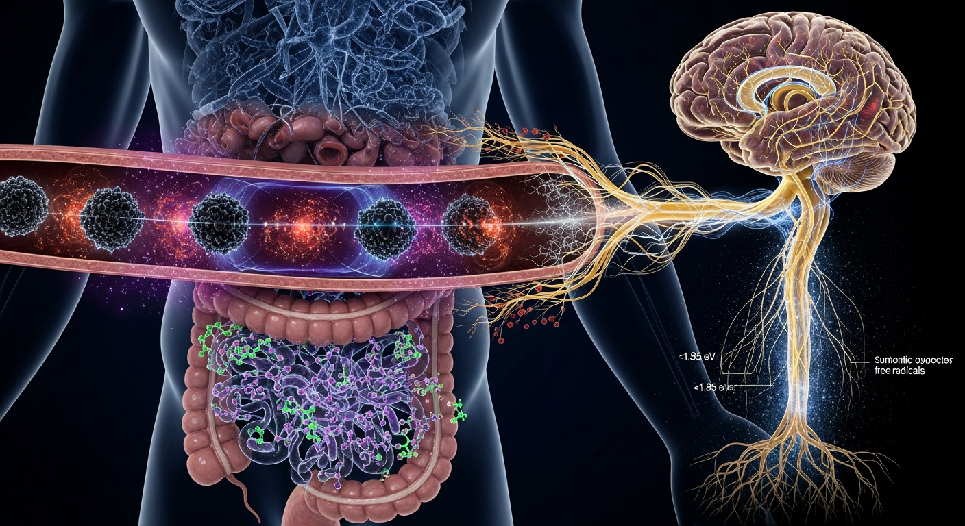
Eumelanin operates with a semiconductor-like bandgap of approximately ~1.85 eV and possesses a remarkable capacity for hydration-dependent proton conductivity. In the highly dynamic and chemically volatile environment of the gut, digestion generates an immense load of reactive oxygen species (ROS) and free radicals. Melanin is singularly equipped to manage this oxidative stress. Because of its highly conjugated planar structure, melanin functions as an electron sink. It stabilizes unpaired electrons, existing continually as a stable free radical—a state easily detectable via Electron Paramagnetic Resonance (EPR) spectroscopy.
Furthermore, the enteric nervous system must continuously process and integrate mechanical, chemical, and osmotic data to drive peristalsis and mucosal secretion. Current biophysical models suggest that melanin in these tissues may go beyond passive ROS scavenging. Given its capacity to transport both electrons and protons, enteric melanin may facilitate localized charge transfer within the extracellular matrix, protecting the delicate membrane potential (Vmem) of enteric neurons from disruption by luminal toxins.
Microbial Modulation of Aromatic Precursors
The biogenesis of melanin in both the gut and the brain relies entirely on the availability of aromatic amino acids, primarily tyrosine and tryptophan. The bioavailability of these critical precursors is fundamentally governed by the gut microbiome. Microbes residing in the intestinal lumen possess the enzymatic machinery to upregulate, degrade, or alter the absorption of these amino acids before they ever reach the bloodstream.
In the classic melanin pathway, tyrosine is oxidized by the enzyme tyrosinase into L-DOPA, which is subsequently converted into dopamine, and eventually polymerized into melanin. In the brain, this exact sequence drives the formation of neuromelanin, the dark pigment characteristic of the substantia nigra. Concurrently, the essential amino acid tryptophan is metabolized in the gut into both serotonin (via the serotonin-melanin pathway) and various indole derivatives.
When the gut microbiome is in a state of eubiosis (balance), it ensures a steady supply of tyrosine and tryptophan into systemic circulation, crossing the blood-brain barrier to fuel both neurotransmitter synthesis and neuromelanin formation. However, under conditions of dysbiosis, specific bacterial strains can sequester these amino acids or shunt them toward toxic metabolic byproducts. This microbial modulation dictates the chemical inventory available to the central nervous system, directly linking the ecology of the gut to the brain's capacity to produce both monoamine neurotransmitters and protective neuromelanin.
Braak's Hypothesis and the Neuromelanin Sink
The profound connection between the gut and brain melanin is most starkly illustrated in the pathology of Parkinson's disease. According to Braak's hypothesis, a leading model in neurobiology, the pathogenesis of Parkinson's does not originate in the brain, but rather in the gut. The theory posits that exposure to an unknown pathogen or toxin in the gastrointestinal tract triggers the misfolding of alpha-synuclein proteins in the enteric nervous system. This pathology then physically travels up the vagus nerve, eventually reaching and devastating the neuromelanin-rich substantia nigra.
Neuromelanin is not a passive bystander in this process; it is a critical neuroprotective agent. Pioneering work by Luigi Zecca and others has demonstrated that neuromelanin acts as a high-affinity chelator for heavy metals, particularly iron, forming a paramagnetic complex that prevents iron-mediated oxidative stress in the cytosol. Neuromelanin also acts as a "chemical sink," sequestering unvesiculated, highly reactive cytosolic dopamine and toxic environmental metabolites.
However, if gut permeability increases—often termed "leaky gut"—a systemic influx of lipopolysaccharides (LPS) and heavy metals enters the bloodstream. This chronic barrage accelerates the depletion of neuromelanin's chelating capacity. Once the neuromelanin polymer becomes saturated with iron and toxins originating from the gut, it undergoes a structural phase shift. Instead of quenching free radicals, saturated neuromelanin begins to generate them, eventually leading to the apoptosis of dopaminergic neurons. Therefore, the preservation of brain neuromelanin is inextricably dependent on the barrier integrity and microbiome health of the gastrointestinal tract.
Bioelectric Networks and Quantum Biology
Looking forward, the intersection of melanin research and bioelectricity offers a compelling theoretical framework for understanding systemic cell communication. Research from Michael Levin's laboratory at Tufts University has demonstrated that endogenous bioelectric networks—driven by ion channels and gap junctions—regulate embryogenesis, tissue regeneration, and anatomical computation independently of the central nervous system.
When we introduce melanin's semiconductor properties into the bioelectric model of the gut, new physiological mechanisms become theoretically possible. Because melanin exhibits hydration-dependent proton conductivity and can facilitate quantum tunneling of electrons within its polymeric structure, extracutaneous melanin may act as a solid-state bioelectric transducer.
It is hypothesized that enteric melanin deposits could function as distributed nodes within the gut's bioelectric field, modulating the bioelectric resting potentials ($V_{mem}$) of surrounding epithelial and immune cells. By acting as a localized buffering system for protons and electrons, melanin may help maintain the bioelectric gradients necessary to prevent the aberrant cellular depolarization associated with chronic intestinal inflammation and tumorigenesis. While the precise quantum mechanical behaviors of melanin in vivo are still being mapped, the recognition of melanin as an active participant in bioelectric signaling fundamentally shifts how we understand the architecture of the gut-brain axis.
Key Takeaways
- Extracutaneous melanocytes in the dark environment of the gastrointestinal tract challenge the traditional view of melanin solely as a photoprotective pigment, highlighting its roles in radical scavenging and electron transport.
- The gut microbiome acts as a master regulator of systemic melanin biogenesis by controlling the metabolism and bioavailability of essential aromatic amino acids like tyrosine and tryptophan.
- Neuromelanin in the brain functions as a critical chemical sink that sequesters heavy metals and toxic metabolites, many of which originate from the gastrointestinal tract during states of intestinal hyperpermeability.
- Braak's hypothesis suggests that neurodegenerative conditions associated with neuromelanin depletion, such as Parkinson's disease, likely originate from pathological protein misfolding in the enteric nervous system.
- Melanin's unique biophysical properties, including ~1.85 eV bandgap semiconductor behavior and hydration-dependent proton conductivity, suggest it may play an active role in maintaining the endogenous bioelectric fields of the gut.
References
Braak, H., Rüb, U., Gai, W. P., & Del Tredici, K. "Idiopathic Parkinson's disease: possible routes by which vulnerable neuronal types may be subject to neuroinvasion by an unknown pathogen." Journal of Neural Transmission 110(5), 517-536 (2003). DOI: 10.1007/s00702-002-0808-2.
Zecca, L., Zucca, F. A., Wilma, A., & Sulzer, D. "Neuromelanin of the substantia nigra: a neuronal black hole with protective and toxic characteristics." Trends in Neurosciences 26(11), 578-580 (2003). DOI: 10.1016/j.tins.2003.08.009.
Plonka, P. M., Passeron, T., Brenner, M., Tobin, D. J., Shibahara, S., Thomas, A., Slominski, A., Kadekaro, A. L., Herskovitz, I., Peters, E., Nordlund, J. J., Abdel-Malek, Z., Zouboulis, C. C., Paus, R., Ortonne, J. P., Hearing, V. J., & Schallreuter, K. U. "What are melanocytes really doing all day long…?." Experimental Dermatology 18(9), 799-819 (2009). DOI: 10.1111/j.1600-0625.2009.00912.x.
Meredith, P., & Sarna, T. "The physical and chemical properties of eumelanin." Pigment Cell Research 19(6), 572-594 (2006). DOI: 10.1111/j.1600-0749.2006.00345.x.
Levin, M. "Molecular bioelectricity: how endogenous voltage potentials control cell behavior and instruct pattern regulation in vivo." Molecular Biology of the Cell 25(24), 3835-3850 (2014). DOI: 10.1091/mbc.E13-12-0708.
O'Mahony, S. M., Clarke, G., Borre, Y. E., Dinan, T. G., & Cryan, J. F. "Serotonin, tryptophan metabolism and the brain-gut-microbiome axis." Behavioural Brain Research 277, 32-48 (2015). DOI: 10.1016/j.bbr.2014.07.027.