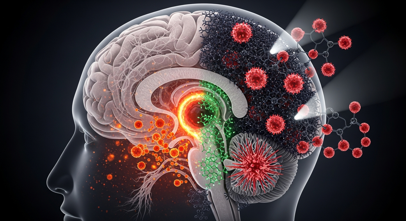
The depigmentation of the substantia nigra is the defining pathological hallmark of Parkinson's disease. This article explores the theoretical framework of targeted melanin precursor therapy—asking whether restoring the brain's internal pigment could fundamentally alter the trajectory of neurodegeneration.
Look at the brain stem of a healthy human adult, and you will see two striking, dark-pigmented streaks. This is the substantia nigra—Latin for "black substance." The deep coloration comes from neuromelanin, a specialized, complex polymer distinct from the eumelanin and pheomelanin found in our skin or hair. For decades, pathologists have known that in the brains of patients with Parkinson's disease, this black substance fades to a pale ghost of its former self. The progressive loss of neuromelanin-containing dopaminergic neurons is the mechanical engine driving the disease's devastating motor symptoms.
But what if this depigmentation is not merely a terminal consequence of cellular death, but a reversible vulnerability? What if, instead of only replacing the dopamine these dying cells once produced, we could physically restore their protective melanin shielding? Imagine a therapeutic intervention designed to "repaint" the substantia nigra—delivering targeted melanin precursors to synthesize functional neuromelanin in surviving neurons, or clearing dysfunctional pigment to rebuild it anew. By shifting our focus from neurotransmitters to the biophysics of neuro-pigmentation, we may uncover a novel method to halt neurodegeneration in its tracks.
The Science We Know
To understand how we might restore neuromelanin, we must first understand what it actually does. Historically dismissed as a metabolic waste product—a mere biological exhaust from dopamine synthesis—neuromelanin is now recognized as a highly sophisticated, neuroprotective sponge.
Neuromelanin is synthesized through the slow oxidation of cytosolic dopamine. When dopamine is not properly packaged into synaptic vesicles, it is highly volatile. Unbound dopamine rapidly autoxidizes into dopamine quinones, generating dangerous reactive oxygen species (ROS) that can tear cellular structures apart. To survive this constant oxidative threat, the neuron converts these volatile quinones into a stable, insoluble polymeric pigment: neuromelanin.
Extensive biophysical profiling, spearheaded by researchers like Luigi Zecca at the Italian National Research Council, has revealed that neuromelanin is a structurally complex matrix of melanin, lipids, and proteins housed within autophagic vacuoles. Because of its unique structure—which features stable free radicals detectable by electron paramagnetic resonance (EPR) spectroscopy—neuromelanin boasts exceptional chelating properties. It acts as a black hole for toxic heavy metals. A healthy neuromelanin organelle can securely bind vast quantities of redox-active transition metals, particularly iron ($Fe^{3+}$), locking them away where they cannot trigger oxidative damage.
However, this biological defense system has a double edge. As dopaminergic neurons age and eventually die, their accumulated neuromelanin is released into the extracellular space. David Sulzer and colleagues have demonstrated that extracellular neuromelanin is highly immunogenic. It activates local microglia, triggering a chronic inflammatory cascade that accelerates the death of neighboring neurons. The very shield that protected the neuron from within becomes a weapon once the cell wall ruptures.
The Possibility
If neuromelanin acts as an essential intracellular buffer against oxidative stress, and if Parkinson's progression is exacerbated by the failure of this buffering system, then therapeutically optimizing neuromelanin in early-stage patients presents a compelling frontier.
Standard Parkinson's therapy relies on L-DOPA, a precursor that transiently boosts dopamine levels to manage motor symptoms. What if we engineered an analogous precursor therapy specifically designed to optimize the neuromelanin pathway? We could hypothesize a targeted delivery of modified indole derivatives—such as stabilized 5,6-dihydroxyindole (DHI) or bespoke dopamine analogues—that readily cross the cell membrane and polymerize into a benign, high-capacity melanin matrix without generating the toxic intermediate quinones associated with natural dopamine oxidation.
Through deductive reasoning: If a synthetic or heavily modified biological precursor could seamlessly integrate into the lysosomes of surviving dopaminergic neurons, it could theoretically provide an immediate, renewed sink for toxic iron and unvesicularized dopamine. This would restore the intracellular redox balance.
An alternative speculative approach involves nanomedicine. Rather than forcing the neuron to synthesize new pigment, researchers might deliver synthetic melanin-like nanoparticles (SMNPs) directly to the substantia nigra. These biocampatible nanoparticles, constructed with an optimized ~1.85eV bandgap to harmlessly dissipate energy, could be functionalized to mimic neuromelanin's iron-chelating properties. By artificially repigmenting the environment with synthetic melanin, we could offload the metabolic burden from the struggling neurons, effectively outsourcing the heavy lifting of iron chelation and radical scavenging to applied biophysics.
Challenges and Unknowns
While intellectually enticing, neuromelanin restoration faces profound physiological hurdles that must be rigorously addressed to maintain scientific viability.
The most pressing challenge is the threshold of toxicity. Landmark research from Miquel Vila's laboratory has demonstrated a critical nuance: continuously increasing neuromelanin production is not universally beneficial. Their work revealed that when neuromelanin accumulates beyond a specific cellular threshold, it physically compromises the neuron's architecture, interfering with lysosomal function and ultimately triggering premature senescence. Therefore, a successful precursor therapy cannot simply be a matter of "more is better." It must be a highly calibrated intervention that balances pigment synthesis with effective lysosomal clearance.
Furthermore, we lack a complete understanding of how neuromelanin synthesis is fundamentally regulated in the human brain. While peripheral melanin (in the skin) is enzymatically driven by tyrosinase, the presence and role of tyrosinase in the substantia nigra remains heavily contested in the literature. If neuromelanin formation is primarily a non-enzymatic autoxidation process, controlling the rate of polymerization using synthetic precursors without accidentally sparking a storm of reactive oxygen species will require unprecedented pharmacological precision.
Finally, the blood-brain barrier (BBB) remains a formidable obstacle. Delivering complex precursor molecules or melanin nanoparticles directly to the pars compacta of the substantia nigra without systemic off-target effects requires advanced targeted liposomal or viral-vector delivery systems that are only now beginning to emerge in clinical trials.
The Path Forward
Moving neuromelanin targeted therapy from speculation to clinical reality demands a highly specific research roadmap.
First, the field must heavily leverage advanced in vitro models. We now have the capability to generate neuromelanin-producing human neurons from induced pluripotent stem cells (iPSCs). These organoid models provide an ideal testing ground for synthetic melanin precursors. Researchers must track the uptake of targeted indoles, observing whether these precursors successfully package into functional autophagic vacuoles or if they aberrantly polymerize in the cytoplasm.
Second, the biophysics of engineered melanin nanoparticles requires deep exploration. Studies must investigate whether SMNPs can be safely absorbed by microglia to neutralize extracellular iron without triggering the inflammatory cascades typically associated with naturally released biological neuromelanin.
The ultimate goal is not to resurrect dead tissue, but to armor the surviving neural architecture. By treating melanin not merely as a biomarker of disease, but as an active, druggable bioelectric and structural target, the Quantum Melanin Research Foundation envisions a future where Parkinson's therapy extends beyond replacing lost signals. The path forward lies in understanding the quantum mechanical and biochemical brilliance of the brain's internal pigments—and learning how to safely replenish the dark matter of the human mind.
Key Takeaways
- Neuromelanin is a structurally complex, neuroprotective polymer in the substantia nigra that safeguards neurons by chelating toxic transition metals like iron and sequestering volatile cytosolic dopamine.
- The progression of Parkinson's disease is intrinsically linked to the loss of these pigmented neurons, resulting in an extracellular release of neuromelanin that triggers severe microglial inflammation.
- Speculative Application: Developing targeted melanin precursors or synthetic melanin-like nanoparticles (SMNPs) could theoretically restore the intracellular buffering capacity of surviving neurons.
- Any therapeutic intervention must carefully navigate cellular thresholds; established research shows that over-accumulation of neuromelanin can disrupt lysosomal function and induce cellular senescence.
- Future research must prioritize human iPSC-derived neuronal models to safely map the precise pharmacokinetics of synthetic melanin precursors before moving to clinical applications.
References
- Zecca, L., Zucca, F. A., Wilma, A., & Sulzer, D. "Neuromelanin of the substantia nigra: a neuronal black hole with protective and toxic characteristics." Trends in Neurosciences 26(11), 578-580 (2003). DOI: 10.1016/j.tins.2003.08.009
- Sulzer, D., Bogulavsky, J., Larsen, K. E., Behr, G., Karatekin, E., Kleinman, M. H., ... & Zecca, L. "Neuromelanin biosynthesis is driven by excess cytosolic catecholamines not accumulated by synaptic vesicles." Proceedings of the National Academy of Sciences 97(22), 11869-11874 (2000). DOI: 10.1073/pnas.97.22.11869
- Carballo-Carbajal, I., Francardo, V., Burbulla, L. F., Ito, K., Peñuelas, N., Vila, M., & Krainc, D. "Brain tyrosinase overexpression implicates age-dependent neuromelanin production in Parkinson's disease pathogenesis." Nature Communications 10(1), 973 (2019). DOI: 10.1038/s41467-019-08858-y
- Zucca, F. A., Segura-Aguilar, J., Ferrari, E., Muñoz, P., Paris, I., Sulzer, D., ... & Zecca, L. "Interactions of iron, dopamine and neuromelanin pathways in brain aging and Parkinson's disease." Progress in Neurobiology 155, 96-119 (2017). DOI: 10.1016/j.pneurobio.2015.09.012
- Double, K. L., Zecca, L., Costi, P., Mauer, M., Griesinger, C., Ito, S., ... & Riederer, P. "Structural characteristics of human substantia nigra neuromelanin and synthetic dopamine melanins." Journal of Neurochemistry 75(6), 2583-2589 (2000). DOI: 10.1046/j.1471-4159.2000.0752583.x
- Liu, Y., Ai, K., & Lu, L. "Polydopamine and its derivative materials: synthesis and promising applications in energy, environmental, and biomedical fields." Chemical Reviews 114(9), 5057-5115 (2014). DOI: 10.1021/cr400407a