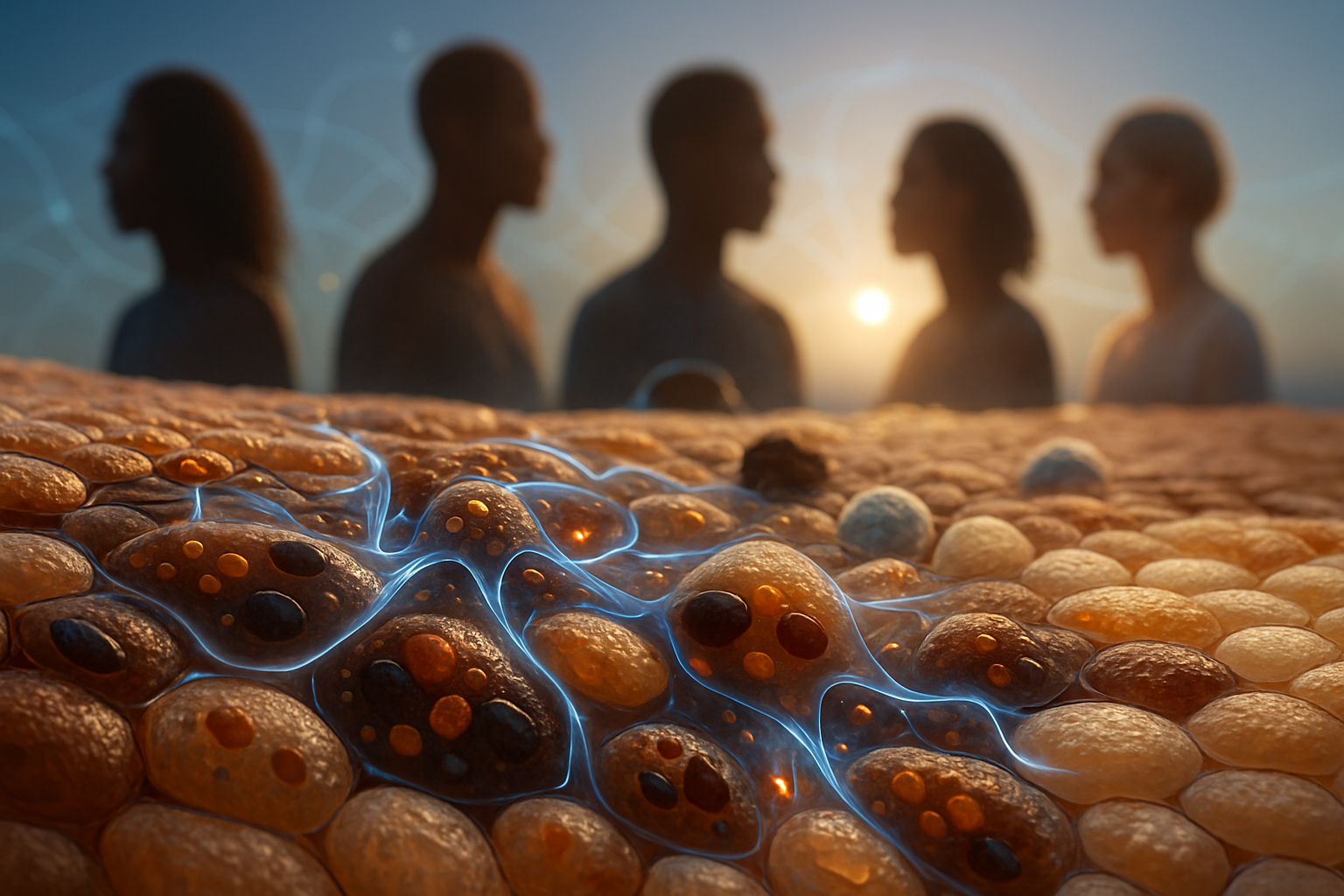
Introduction: The Intersection of Biophysics and Health
Could genetically influenced differences in melanin composition and organization alter the electrical and redox behavior of pigment cells, potentially explaining variations in disease susceptibility among individuals or populations?
This question warrants a detailed exploration that separates established mechanisms from plausible but untested extensions. Melanin bioelectrics refers broadly to the electrical, electrochemical, and redox properties of melanin-containing biological systems. It is not a single mechanism, and the term should not imply that melanin functions as a conventional wire or as an independent signaling organ. At least three partially overlapping processes are relevant:
- Redox activity: Melanin can reversibly accept and donate electrons, influencing radical dissipation, metal-ion chemistry, and oxidative reactions.
- Electronic and protonic conductivity: Hydrated or synthetic melanin preparations can facilitate charge transport, with observed conductivities varying over many orders of magnitude depending on composition and measurement conditions.
- Tissue-scale bioelectric signaling: Cells generate membrane potentials, transepithelial potentials, and extracellular electric fields that can influence migration, proliferation, repair, and developmental patterning.
These levels must be distinguished carefully. A redox-active pigment can alter the lifetime of reactive oxygen species without carrying a physiologically meaningful current across a tissue. A melanin film can exhibit measurable conductivity between electrodes without forming a continuous electrical pathway through intact melanosomes. Similarly, a wound-associated electric field may be generated primarily by ion pumps, epithelial junctions, and extracellular resistance rather than by pigment.
The strongest established connection between melanin biology and human disease involves ultraviolet radiation (UVR) photobiology, oxidative damage, and melanoma. The relative abundance and organization of eumelanin and pheomelanin, influenced by genes such as MC1R, can affect UV absorption, reactive intermediates, DNA damage, and repair responses. These mechanisms plausibly contribute to differences in melanoma susceptibility, especially when considered alongside total pigmentation, nevus burden, UV exposure, immune surveillance, age, and inherited cancer-risk variants.
A direct connection between melanin conductivity and population-level differences in infection, wound repair, systemic disease, or generalized drug response has not been demonstrated. Such a claim would require evidence that:
- melanin carries charge at relevant rates under physiological conditions;
- the current or redox flux is spatially organized;
- the flux is coupled to a cellular signaling pathway;
- genetic or biochemical differences alter that flux in vivo; and
- the resulting change predicts a clinically measurable outcome after accounting for confounding variables.
This is a demanding standard, but it is scientifically useful. The Quantum Melanin Research Foundation (QMRF) views the subject as a biophysical frontier: identifying measurable molecular properties that could connect genotype, environment, tissue physiology, and clinical outcomes. The central thesis is therefore significant but cautious. Melanin composition already influences disease risk through established photochemical and redox pathways. A direct role for melanin-mediated charge transport remains a testable hypothesis rather than an established feature of human physiology.
The Biophysics of Melanin: From Pigment to Redox Activity
Understanding Melanin as a Chemically Diverse Material
Melanin is not a singular molecule with a fixed molecular weight, crystal lattice, or universally defined electronic structure. It is better understood as a family of oxidized and reduced aromatic oligomers that assemble into heterogeneous supramolecular particles. Their properties depend on precursor chemistry, oxidation state, hydration, particle size, aggregation, metal binding, protein association, pH, and preparation history.
Eumelanin is generally associated with indole-derived units formed through the oxidation and subsequent reactions of tyrosine and dopa. Its structure includes mixtures of quinone, hydroquinone, semiquinone, indole, and indole-quinone-like groups. The exact distribution of these groups varies across tissues and experimental preparations. Eumelanin's broad absorption across ultraviolet and visible wavelengths allows it to dissipate absorbed energy efficiently, often converting it into heat through ultrafast nonradiative relaxation. It can also interact with free radicals and redox-active metals.
Pheomelanin contains sulfur-bearing benzothiazine and benzothiazole-related structures produced when cysteine or other sulfur-containing compounds participate in the melanogenic pathway. Pheomelanin can absorb light, but its photochemical behavior differs from eumelanin. Under some conditions, it can participate in reactions that generate reactive oxygen species or other radical intermediates. The biological effect depends on illumination wavelength, oxygen concentration, local redox state, metal content, and the molecular organization of the pigment.
The distinction between the two classes is useful but not absolute. Natural melanosomes can contain mixtures of eumelanin and pheomelanin, and their chemical environment is shaped by proteins, lipids, membrane-associated transporters, and the melanosome's internal pH. Melanin is synthesized through enzymatic pathways involving tyrosinase and related proteins, and the melanosome is not simply a container filled with pigment. It is a specialized organelle whose architecture may determine how efficiently radicals, photons, protons, and metal ions interact with pigment.
A small change in composition can have several consequences simultaneously. For example, increasing pheomelanin content may alter:
- the wavelengths and efficiency of light absorption;
- the availability of sulfur-containing redox sites;
- the balance between radical scavenging and radical generation;
- the binding of metal ions;
- the oxidation state distribution of pigment;
- the amount of oxidative stress transmitted to nearby membranes or nuclei.
These effects should not be interpreted as a simple “protective versus harmful” binary. Eumelanin can undergo oxidative chemistry as well, and pheomelanin can contribute to light absorption and radical quenching under some conditions. The biologically relevant variable may be the entire redox environment rather than pigment class alone.
Redox Activity and Charge Transport Explained
Melanin's redox activity arises from chemically interconvertible functional groups. In a simplified representation:
[ \text{quinone} + 2H^+ + 2e^- \rightleftharpoons \text{hydroquinone} ]
Semiquinone-like radicals can occur between these states:
[ \text{quinone} + H^+ + e^- \rightleftharpoons \text{semiquinone} ]
These equations do not describe a single molecular reaction occurring uniformly throughout a melanosome. They represent families of related redox states distributed across an irregular polymeric and oligomeric network. Their practical significance is that melanin can act as a redox buffer: it may accept electrons from some species and donate them to others, depending on local potential, pH, oxygen availability, and the presence of metal ions.
A redox buffer does not necessarily consume radicals irreversibly. If a pigment accepts an electron and later transfers it to oxygen, a metal ion, or another acceptor, it may redistribute oxidative equivalents rather than eliminate them. This distinction matters when interpreting claims that melanin “neutralizes” oxidative stress. The outcome depends on reaction rates and pathways.
Proposed charge-transport mechanisms include:
- Electron hopping: An electron moves between spatially separated redox-active sites. In a disordered material, this process may occur through thermally assisted hopping rather than through a delocalized conduction band.
- Proton conduction: Protons move through hydrogen-bonded water networks and protonatable functional groups. This process can increase substantially with hydration.
- Mixed ionic/electronic transport: Electrons and ions move together, with changes in one influencing the other. This is common in soft, hydrated electrochemical materials.
- Interfacial charge transfer: Electrodes, water layers, counterions, and surface chemistry can dominate the measured current.
- Redox-mediated transport: A redox-active site accepts an electron, changes state, and transfers an electron to a neighboring site. The measured current may therefore reflect sequential chemical reactions rather than the movement of free electrons through a uniform solid.
The distinction between electron and proton transport is especially important. A hydrated melanin preparation may conduct more effectively because water increases ion mobility, changes dielectric screening, and permits proton exchange. An experiment that reports increased current after hydration does not by itself establish that electrons are moving through the pigment in the same way as they would in a dry film.
A useful worked example illustrates the scale of the issue. Suppose a melanin film has a conductivity of (10^{-7}) S/cm, an applied electric field of (1) V/cm, and a cross-sectional area of (0.01) cm². Ohm's law gives:
[ J = \sigma E = (10^{-7}\ \text{S/cm})(1\ \text{V/cm}) = 10^{-7}\ \text{A/cm}^2 ]
The total current would be:
[ I = JA = (10^{-7}\ \text{A/cm}^2)(0.01\ \text{cm}^2) = 10^{-9}\ \text{A} ]
or approximately 1 nanoampere. That is readily measurable with laboratory instrumentation. However, this calculation does not establish that the same current flows through a melanosome, across a cell, or through intact skin. The geometry, electrode contact, hydration, voltage, and path length are all different. The example demonstrates measurement feasibility, not physiological significance.
Clarifying Conductivity Measurements
Reported conductivity values for melanin span approximately (10^{-10}) to (10^{-5}) S/cm, although values outside this range may appear depending on the material and method. This range covers five orders of magnitude. Such variation is not necessarily evidence of contradictory science; it reflects the fact that different experiments may measure materially different substances under different conditions.
Important variables include:
- Hydration: Water can increase ionic and protonic transport, alter molecular spacing, and modify redox kinetics.
- Relative humidity: A film measured at low humidity may behave very differently from the same film at 80–90% relative humidity.
- Film thickness: Contact resistance and nonuniform hydration can distort apparent conductivity.
- Electrode material: Gold, platinum, carbon, and other electrodes can interact differently with redox-active pigments.
- Applied voltage: At higher fields, nonlinear behavior, charge injection, electrochemical reactions, or heating may occur.
- Frequency: Alternating-current impedance measurements can distinguish, imperfectly, between bulk resistance, interfacial capacitance, and electrode polarization.
- Pigment source: Synthetic eumelanin, sepia melanin, enzymatically generated pigment, and human melanosomal pigment are not interchangeable.
- Oxidation state and pH: The number and accessibility of redox-active sites can change with chemical treatment.
- Particle organization: Aggregates, films, and dispersed particles create different transport pathways.
Historical research, including work associated with McGinness and later device-oriented studies such as those by Bothma and colleagues, demonstrated that melanin preparations can support measurable electrical transport and can display unusual switching or hydration-dependent behavior. These findings established that melanin is electrically active in a materials-science sense.
The translational question is narrower: does melanin carry charge in intact human tissue at a magnitude and organization capable of influencing cell behavior?
Addressing this question requires measurements in or near native structures. A suspension of melanin particles, a compressed powder, and a hydrated thin film may each generate a current, but their results cannot automatically be generalized to a melanosome embedded in a keratinocyte. In living skin, melanosomes are surrounded by membranes, cytoplasm, ions, proteins, and extracellular matrix. Their electrical properties would be measured in parallel with many other conductors and capacitors.
A physiologically meaningful demonstration would need at least three components:
- a direct measurement of charge or redox flux associated with melanosomes under controlled conditions;
- manipulation of melanin chemistry without broadly damaging the cell; and
- a reproducible change in a downstream cellular process, such as calcium signaling, migration, oxidative stress, or repair.
Using “Band Gap” Language With Caution
Reported energy scales for melanin preparations often fall near 1.5–2.0 eV, but these values should not be treated as a single universal band gap. In ordered crystalline semiconductors, the band gap separates well-defined valence and conduction bands. Melanin is chemically and structurally disordered. Its optical absorption may reflect overlapping transitions among many molecular sites, localized states, aggregates, and charge-transfer complexes.
A more careful description uses concepts such as:
- localized electronic states;
- energetic disorder;
- hopping barriers;
- density of redox-active sites;
- hydration-dependent mobility;
- interfacial polarization;
- broad optical absorption.
A reported optical energy scale might be obtained from an absorption spectrum, while an electrical activation energy might be inferred from temperature-dependent conductivity. These are not necessarily the same quantity. For example, an optical transition near 1.8 eV does not mean that an electron can travel through a biological melanosome with an energy barrier of exactly 1.8 eV.
The key principle is that laboratory conductivity demonstrates potential capability, not inherent physiological function. The relevant physiological comparison is not whether melanin can conduct under any conditions, but whether it conducts faster or more selectively than the surrounding cytoplasm, organelle membrane, extracellular fluid, and tissue matrix—and whether that difference changes biology.
Genetic and Biochemical Pathways Linking Melanin to UV-Related Conditions
Polygenic Nature of Pigmentation
Human pigmentation is polygenic. Genes including MC1R, TYR, TYRP1, OCA2, SLC24A4, SLC45A2, KITLG, and others influence melanocyte development, melanosome formation, pigment synthesis, intracellular transport, and pigment distribution. Their effects are modified by ancestry, hormonal state, age, inflammation, UV exposure, and local tissue environment.
MC1R is particularly important because signaling through the melanocortin-1 receptor tends to favor eumelanin production over pheomelanin production. Variants that reduce receptor signaling can be associated with red or blond hair, lighter skin, freckling, and altered melanoma susceptibility. However, genotype is not a direct measurement of pigment chemistry. Two people carrying the same MC1R variant may differ in total melanin content, pheomelanin fraction, tanning response, nevus count, UV exposure, and DNA repair capacity.
Similarly, visible pigmentation is an imperfect proxy for melanin composition. Skin reflectance can estimate optical properties, but it does not by itself reveal:
- the eumelanin-to-pheomelanin ratio;
- the oxidation state of the pigment;
- melanosome size and density;
- pigment distribution within the epidermis;
- the rate of melanosome degradation;
- redox activity under illumination;
- the degree of metal binding or protein association.
A mechanistic study should therefore measure biochemical and biophysical phenotypes directly whenever possible. Genotype can define a causal or predictive variable, but it should not substitute for measuring the actual pigment state.
Established Photobiological Functions
Melanin absorbs ultraviolet radiation and helps reduce the amount of UV energy reaching nuclear DNA. Melanosomes are distributed within keratinocytes, often forming supranuclear caps or protective arrangements that create a physical and optical barrier. Their protective action depends on pigment concentration, particle size, position, persistence, and chemical composition.
UVB, approximately 280–315 nm, is strongly associated with direct DNA photoproducts such as cyclobutane pyrimidine dimers and 6-4 photoproducts. UVA, approximately 315–400 nm, penetrates more deeply and contributes substantially to oxidative stress through photosensitization and reactive oxygen species. Melanin can attenuate both types of exposure, but attenuation is not equivalent across wavelengths or pigment chemistries.
A simplified attenuation model is:
[ I(z)=I_0 e^{-\alpha z} ]
where (I_0) is incident intensity, (I(z)) is intensity after traveling distance (z), and (\alpha) is an effective absorption coefficient. In real skin, scattering, reflection, melanosome geometry, and cellular organization complicate this expression. Nevertheless, it illustrates why pigment distribution matters. A high concentration of pigment in a thin supranuclear layer may protect DNA differently from the same total amount dispersed throughout the cytoplasm.
The evolutionary relationship between pigmentation and geographic UV exposure is well established at the population level, but it should not be misinterpreted as a complete explanation of disease risk. UV exposure varies substantially within every population, and individual behavior, occupation, latitude, altitude, clothing, sun-protection practices, immune status, and medical surveillance all influence outcomes.
Eumelanin, Pheomelanin, and Oxidative Stress
Eumelanin-rich systems generally provide strong broadband absorption and can dissipate excitation energy rapidly. Their redox-active sites can interact with free radicals and metal ions. Pheomelanin-rich systems may display different sulfur chemistry and can, under illumination, contribute to oxidative reactions. The balance between these processes is affected by oxygen tension, pH, antioxidant availability, and pigment oxidation state.
A worked example helps explain why pigment quantity alone is insufficient. Imagine two melanocyte-derived systems with equal total melanin mass:
- System A contains 80% eumelanin and 20% pheomelanin.
- System B contains 40% eumelanin and 60% pheomelanin.
If both receive the same UVA dose, their total optical absorption might be similar, but the chemical consequences may differ. System B could generate a larger oxidative signal if its pheomelanin-associated pathways produce more reactive intermediates under those conditions. System A might dissipate excitation more efficiently or provide different radical-buffering behavior. Yet the result could reverse or diminish in a low-oxygen environment, in the presence of antioxidants, or after changes in pigment oxidation state.
This is why causal separation is difficult. Pheomelanin-rich phenotypes often coincide with lighter skin, reduced tanning, freckling, and different UV exposure patterns. An observed increase in melanoma risk may therefore reflect several linked pathways rather than one isolated property of pheomelanin.
Melanoma: The Strongest Disease Link
Melanoma provides the most compelling disease context for studying melanin chemistry. UV radiation can cause direct DNA lesions, oxidative base damage, mitochondrial stress, inflammation, and alterations in signaling pathways that regulate melanocyte survival and proliferation. Melanocytes are particularly relevant because they synthesize pigment while residing in a tissue exposed to solar radiation.
One important finding is melanin-associated chemiexcitation. In experimental systems, UV-induced chemical reactions can generate electronically excited states that persist after the initial illumination event. These excited states may transfer energy to DNA and produce delayed cyclobutane pyrimidine dimers. The effect is especially relevant in systems containing pheomelanin or high oxidative stress, although the magnitude depends on experimental conditions.
The timing is mechanistically important. Direct UV photoproducts form during irradiation, whereas chemiexcitation-associated damage can continue afterward. If a substantial fraction of damage occurs during a post-illumination window, then antioxidant state, temperature, oxygen availability, and melanin composition may influence risk even after the UV source is removed.
Melanoma risk is nevertheless multifactorial. Important variables include:
- inherited variants in pigmentation and cancer-related genes;
- total and intermittent UV exposure;
- history of sunburn;
- number and type of melanocytic nevi;
- age and immune function;
- family history;
- somatic mutations acquired by melanocytes;
- DNA repair and cell-cycle responses.
These findings establish a meaningful link between melanin chemistry and disease susceptibility but do not prove that melanin conductivity is responsible. A direct electrical mechanism would require evidence that charge transport alters, for example, DNA repair, calcium signaling, mitochondrial function, or melanocyte survival in a way that is independent of ordinary photochemical redox reactions.
Bridging the Gap: From Isolated Pigment to Functioning Tissue
Melanin's biological function occurs in melanosomes, not in purified pigment alone. A melanosome contains a polymeric pigment matrix enclosed by a membrane and associated with structural and enzymatic proteins. Its lumenal pH, ionic composition, and maturation state influence melanogenesis. Mature melanosomes are transferred from melanocytes to keratinocytes, where they persist and are eventually degraded.
This organization creates several possible barriers to electrical coupling. The melanin matrix may be physically separated from the cytosol by a membrane. The membrane may be electrically insulating or selectively permeable. Counterions may be required to maintain local charge neutrality. Water content may differ between the pigment matrix and surrounding cytoplasm. Any current generated by melanin would therefore have to be coupled to nearby conductive pathways.
Evidence spans three scales:
- Molecular scale: Electron transfer, proton exchange, radical buffering, metal binding, and chemical excitation.
- Material scale: Measurable transport in synthetic films, powders, dispersions, and hydrated preparations.
- Tissue scale: Bioelectric fields and currents associated with epithelial barriers, wound edges, ion pumps, gap junctions, and extracellular matrices.
At the tissue scale, epithelial wounds can generate endogenous electric fields. Measurements in experimental epithelia have commonly reported field strengths on the order of tens to hundreds of millivolts per millimeter, although values depend on species, tissue type, wound geometry, measurement location, and time after injury. These fields can influence directional migration, or galvanotaxis, in keratinocytes and other cell types. The major established sources include transepithelial potential differences, ion-selective transport, exposed wound surfaces, and current flow through extracellular pathways.
A simple estimate illustrates why tissue-level signals are plausible without invoking melanin. If a wound-associated field is (100) mV/mm, then across a (100\ \mu\text{m}) cell:
[ \Delta V = E d ]
[ \Delta V = (100\ \text{mV/mm})(0.1\ \text{mm}) = 10\ \text{mV} ]
A 10 mV difference across the length of a cell can influence voltage-sensitive molecular processes, membrane transport, and polarized signaling. This field can arise from epithelial ion transport alone. Melanin might modulate it only if pigment-associated conductivity or charge storage measurably changes current distribution.
The central gap lies in measuring these properties under physiological conditions. A convincing study would need to distinguish:
- current through melanin from current through the surrounding electrolyte;
- proton transport from electron transport;
- electrode artifacts from biological signals;
- pigment-dependent effects from changes in cell viability;
- optical or redox effects from genuinely electrical effects.
Appropriate tools could include impedance spectroscopy, scanning electrochemical microscopy, redox-sensitive fluorescent probes, genetically encoded voltage or calcium indicators, Raman or electron-paramagnetic-resonance measurements, and high-resolution imaging of melanosome organization. The most informative experiments would combine these methods rather than relying on a single conductivity number.
A Proposed Study: Analyzing Melanin Bioelectrics and Skin Recovery
Envision a study examining whether pigment chemistry and bioelectric properties influence skin recovery after controlled injury. The design should prioritize individual biochemical phenotypes rather than assigning biological properties to broad demographic categories.
Participants could be characterized using:
- quantitative skin reflectance at several wavelengths;
- hair or skin pigmentation phenotype;
- targeted genotyping of pigmentation-related variants;
- melanin density and melanosome morphology from noninvasive imaging or ethically justified tissue samples;
- biochemical estimates of eumelanin and pheomelanin markers;
- baseline antioxidant capacity and inflammatory status;
- UV exposure history and sun-protection behavior.
For pigmentation chemistry, one possible approach would use degradation products associated with eumelanin and pheomelanin. Pyrrole-2,3,5-tricarboxylic acid is commonly used as an eumelanin-associated marker, while sulfur-containing degradation products such as benzothiazine-related units can inform pheomelanin content. These measurements are imperfect and require standardized sample collection, normalization to tissue mass or protein, and careful treatment of oxidative artifacts.
Electrical measurements should be performed at several levels. First, purified or isolated melanin preparations from the same participants could be examined under controlled humidity, pH, ionic strength, and temperature. Second, melanocytes and keratinocytes could be studied in culture, with melanosome content manipulated while maintaining viability. Third, reconstructed epidermis or organotypic skin could be used to measure wound-edge potentials and currents.
A controlled wound model might use a standardized scratch or micro-injury in a reconstructed epidermis. The primary outcomes could include:
- wound closure after 6, 12, 24, and 48 hours;
- directionality and velocity of keratinocyte migration;
- wound-edge electric field strength;
- intracellular calcium transients;
- reactive oxygen species levels;
- mitochondrial membrane potential;
- cytokines such as IL-1β, IL-6, TNF, and TGF-β;
- barrier restoration measured by transepithelial electrical resistance;
- melanocyte survival and pigment redistribution.
A useful experimental matrix would include at least four conditions:
- pigment-deficient cells or tissues;
- eumelanin-enriched systems;
- pheomelanin-enriched systems;
- matched pigment systems in which redox chemistry is altered without changing total pigment mass.
The fourth condition is crucial. If an effect occurs only when pigment amount changes, it may reflect optical shielding or physical structure. If it persists when total pigment is held constant but redox state is manipulated, a chemical mechanism becomes more likely. If a selective melanin redox or transport inhibitor abolishes the effect while cell viability remains intact, an electrical or electrochemical contribution becomes more plausible.
The experiment should also include an external-field control. Applying a weak, calibrated electric field to pigment-deficient tissues could test whether pigmentation changes the response to a known field. For example, if two tissues experience the same external field but show different migration rates, one could ask whether melanin modifies field sensing, membrane polarization, or redox signaling.
A worked analysis might proceed as follows. Suppose:
- Tissue A contains 2.0 arbitrary units of eumelanin marker per milligram of protein.
- Tissue B contains 1.0 unit of eumelanin marker and 1.0 unit of pheomelanin marker.
- Both tissues show the same wound-edge field: (80) mV/mm.
- Tissue A closes 90% of the wound in 24 hours.
- Tissue B closes 72% in 24 hours and shows a twofold higher oxidative fluorescence signal.
This result would support a difference in repair-associated oxidative biology, but it would not establish conductivity as the cause because the electric fields are identical. If, however, Tissue B showed a different local current distribution despite the same measured macroscopic field, and if that difference tracked with melanosome-associated impedance, a bioelectric contribution would become more credible.
Statistical analysis should account for repeated measurements, donor-to-donor variability, batch effects, baseline pigmentation, UV history, and tissue maturity. A mixed-effects model could treat donor as a random effect and pigment composition, genotype, hydration, and field strength as fixed effects. The study should be powered to detect biologically meaningful changes rather than merely statistically significant differences in fluorescence.
Ethical and interpretive safeguards are essential. Population labels should not be used as mechanistic substitutes for pigment measurements. Human pigmentation traits reflect ancestry, but ancestry is not a direct measure of melanin chemistry, electrical properties, immune function, or clinical prognosis. The scientifically relevant variables are measurable phenotypes and exposures.
Looking Forward: Testing Hypothetical Extensions of Melanin Chemistry
Melanin's participation in redox chemistry and laboratory conductivity suggests several experimentally testable extensions, but each requires a narrowly defined mechanism.
One hypothesis is that melanin could alter local oxidative environments during tissue repair. Melanin-rich organelles might buffer radicals generated by inflammation, or they might amplify oxidative reactions under specific illumination and metal-ion conditions. This can be tested by comparing redox flux, rather than simply measuring total pigment, in matched cell systems.
A second hypothesis is that melanin could influence local charge distribution or impedance. A melanosome might function as a microscopic redox capacitor, storing and releasing charge over time. This would predict measurable frequency-dependent behavior. For example, impedance spectroscopy could determine whether melanin-containing cells exhibit a distinct relaxation time compared with pigment-deficient cells. Such an effect would be more persuasive if it scaled with melanosome number and persisted after controlling for cell size, membrane capacitance, and viability.
A third hypothesis concerns light-triggered charge or proton movement. Melanin absorbs across broad wavelengths, but absorption alone does not establish useful photoconversion. Experiments should compare dark conditions with controlled UVA, visible, and near-infrared exposures, measuring proton flux, redox state, calcium activity, and membrane potential. Thermal controls are necessary because pigment absorption can produce local heating.
A fourth possibility is that pigment chemistry could alter drug response. This is plausible for drugs that bind melanin or undergo redox cycling, but it should not be generalized to all medications. A mechanistic experiment would compare drug accumulation, intracellular free concentration, binding affinity, and cytotoxicity in cells with defined melanin contents. If melanin sequesters a compound, the relevant prediction may be delayed pharmacologic action rather than altered electrical signaling.
Any proposed link to infection would require particular caution. Melanin may affect microbial adhesion, oxidative chemistry, light sensitivity, or local barrier properties, but there is currently no general evidence that pigment conductivity determines susceptibility to infection. Studies should identify a specific organism, tissue site, pigment-dependent biochemical reaction, and measurable outcome.
These hypotheses should be evaluated with perturbation and rescue experiments. Correlation between pigmentation and an outcome is not sufficient. Stronger evidence would come from changing melanin composition, restoring it, altering its oxidation state, or modifying hydration while independently measuring the proposed electrical or redox intermediate.
Key Takeaways and Future Directions
- Melanin is a heterogeneous, redox-active pigment system, not a single uniform molecule. Its behavior depends on oligomer composition, oxidation state, hydration, pH, metals, proteins, and supramolecular organization.
- Eumelanin and pheomelanin differ chemically and photobiologically. Eumelanin generally provides strong broadband absorption and radical-buffering capacity, while pheomelanin-associated sulfur chemistry can contribute to oxidative reactions under particular conditions.
- Melanin can conduct charge in laboratory preparations. Reported conductivities, approximately (10^{-10}) to (10^{-5}) S/cm, vary widely because hydration, electrode interfaces, morphology, and measurement protocol strongly influence results.
- Conductivity is not equivalent to physiological signaling. Demonstrating a current in a film does not prove that intact melanosomes carry a biologically meaningful current through human skin.
- The strongest established disease connection is UV-related damage and melanoma susceptibility. Relevant mechanisms include optical shielding, oxidative stress, pigment composition, chemiexcitation, DNA damage, and interactions with inherited and environmental risk factors.
- Genotype does not equal pigment chemistry. MC1R and other pigmentation genes influence pathways, but direct measurement of eumelanin, pheomelanin, melanosome organization, redox state, and exposure history is necessary for mechanistic conclusions.
- Tissue bioelectricity already has established sources. Wound-associated fields arise principally from epithelial ion transport, membrane potentials, and extracellular current pathways. Melanin's contribution, if present, must be separated from these dominant mechanisms.
- Future studies should integrate molecular, material, cellular, and tissue-scale measurements. Conductivity, impedance, proton flux, redox state, membrane potential, calcium signaling, migration, and clinical outcomes should be analyzed together.
- Population-health interpretations require biochemical precision. Broad racial or ancestral categories should not be treated as proxies for electrical properties or disease mechanisms. Individual-level phenotypes and environmental exposures are more informative and scientifically defensible.
QMRF's biophysical lens supports disciplined exploration rather than premature certainty. Melanin may prove important in a narrow set of conditions involving UV exposure, oxidative stress, hydration, redox-active drugs, or tissue repair. The decisive evidence will come from experiments that connect a defined pigment property to a defined cellular pathway and then to a reproducible physiological outcome. Until such evidence exists, melanin-mediated charge transport should remain a promising hypothesis that complements—not replaces—the established photochemical and genetic explanations of pigmentation-associated disease susceptibility.