Introduction: The Promise and the Measurement Challenge
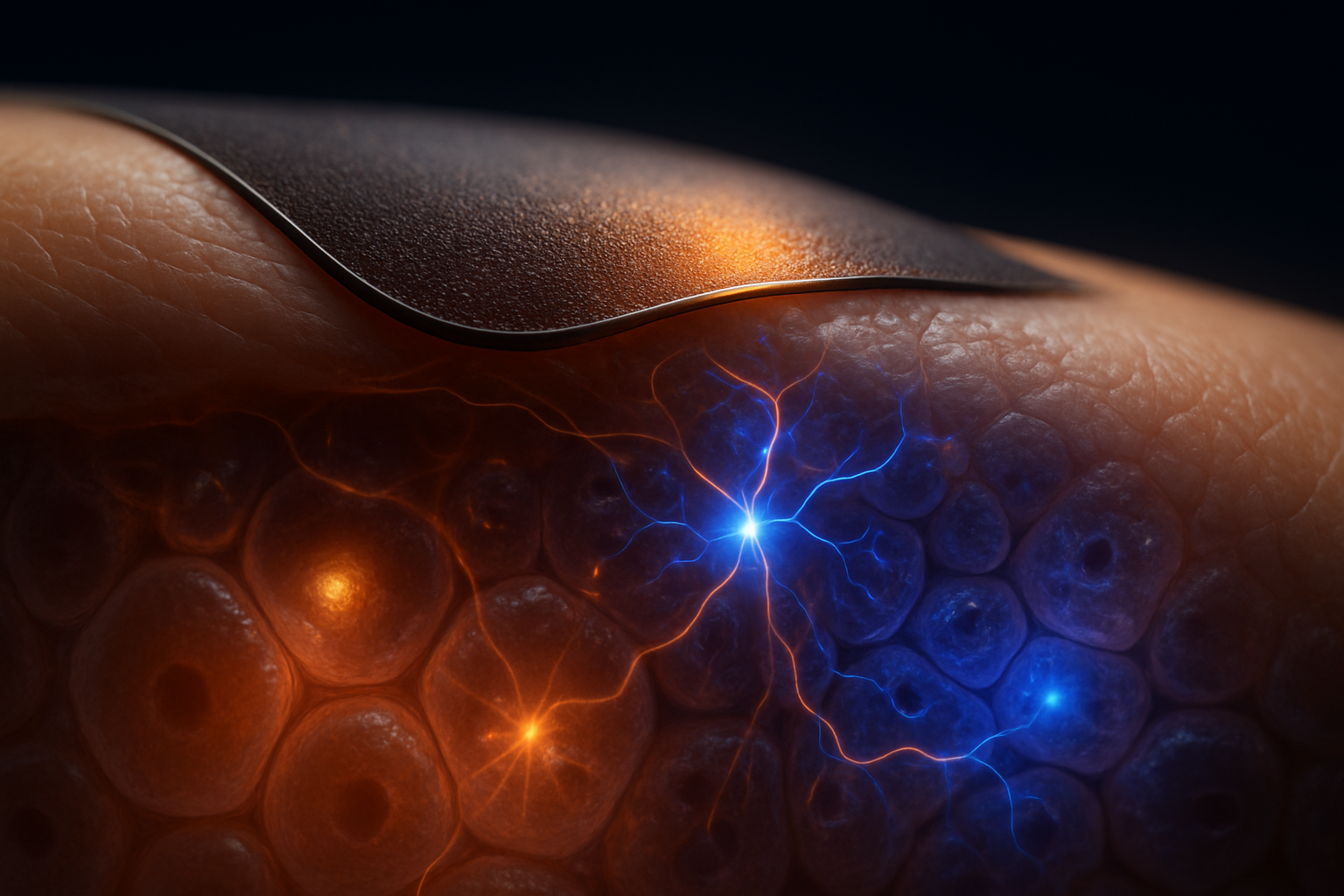
Imagine a soft, biocompatible interface constructed from melanin, capable of detecting the electrical reorganization associated with cancer transformation before a tumor becomes observable or symptomatic. This innovative device would not focus solely on identifying a cancer-specific protein or circulating metabolite. Instead, it would aim to detect a deeper systemic pattern: how transformed cells regulate ions, membrane voltage, redox chemistry, and intercellular electrical communication.
The foundation of this concept is built upon two critical scientific observations. Firstly, cancer and transformed cells often display altered resting membrane potentials, with a trend toward depolarization compared to their non-transformed counterparts. Bioelectric states are not merely passive electrical phenomena; they can actively influence processes such as proliferation, differentiation, migration, invasion, and tissue organization through voltage-sensitive ion channels, calcium signaling, gap junctions, and downstream transcriptional pathways. Secondly, eumelanin is recognized as an electroactive biological material, featuring a remarkable combination of electronic, ionic, protonic, and redox-mediated charge transport.
Despite these promising observations, there remains a clear boundary between established scientific knowledge and speculation. Although cancer-associated bioelectricity is a legitimate research focus and melanin possesses measurable electrochemical properties, no peer-reviewed study to date has demonstrated the ability of a melanin-based wearable to detect a cancer-specific signal in humans, let alone a pre-cancerous signal through intact skin.
The core measurement challenge lies in the disparity between intracellular and extracellular measurements. While a membrane potential is inherently an intracellular, cell-scale phenomenon, a wearable sensor typically measures extracellular, tissue-level activities. Thus, the pivotal question becomes whether a melanin-based interface can register a localized, heterogeneous cancer-associated state and translate it into a consistent signal that is distinguishable from factors such as hydration, pH, inflammation, wound repair, infection, hypoxia, and the normal remodeling of tissues.
Cancer Bioelectricity: Understanding Malignant and Pre-Malignant Transformations
Resting Membrane Potential: A Key Cellular Regulator
The resting membrane potential represents the voltage difference across a cell's membrane, arising from unequal ion concentrations, selective permeability, and active transport mechanisms. The Na⁺/K⁺-ATPase enzyme plays a critical role by exporting sodium ions while importing potassium ions, creating the gradients necessary to generate voltage through ion channels and transporters. Potassium permeability is often essential, as the plasma membrane is typically more permeable to K⁺ at rest.
The equilibrium potential for an ion can be approximated using the Nernst equation:
[ E_{\mathrm{ion}}=\frac{RT}{zF}\ln\left(\frac{[\mathrm{ion}]{\mathrm{out}}}{[\mathrm{ion}]{\mathrm{in}}}\right), ]
with the overall membrane voltage being more accurately represented by the Goldman–Hodgkin–Katz equation. This resting membrane voltage is dynamic, varying with changes in channel opening, transporter expression, cell volume, metabolism, and the extracellular ionic environment.
Many mammalian cells have resting potentials ranging from approximately -60 to -90 mV. In contrast, transformed or cancerous cells frequently exhibit less negative potentials, between -20 and -40 mV. While the comparison between -70 mV in differentiated cells versus -20 mV in cancerous cells is instructive, it should not be viewed as an absolute biological law, as values depend on factors such as tissue type, cell cycle, and cancer subtype.
Ion Channels and Signaling Pathways in Cancer
Malignant transformation may alter the expression or activity of various ion channels and transport systems, including voltage-gated and ligand-gated ion channels, inward-rectifier potassium channels, sodium and calcium channels, and proton transporters. These shifts can affect membrane voltage and, concurrently, alter intracellular chemical dynamics.
Depolarized membranes may modify the driving force for calcium entry, with intracellular Ca²⁺ acting as a second messenger that can impact calmodulin-dependent enzymes, protein kinases, cytoskeletal organization, mitochondrial activity, and transcription factors. Voltage-regulated pathways can intersect with signaling cascades involving MAPK, PI3K–AKT, cell-cycle control, and apoptosis. Conversely, oncogenic signaling can alter channel abundance and pump activity, creating a feedback loop where altered signaling influences voltage, which in turn reinforces the cancer phenotype.
Gap junctions provide a tissue-scale layer of complexity. Connexin-based channels enable ions and small molecules to flow between adjacent cells, and cancer-related changes in gap-junction expression or positioning can electrically isolate subpopulations. Abnormal coupling among tumor cells, stroma, endothelium, and immune cells may further modify local bioelectric domains.
Functionality of Bioelectricity in Cancer
Studies by Yang and Brackenbury, as well as research by Sundelacruz, Levin, and Kaplan, illustrate associations between membrane-potential changes and cellular behaviors such as proliferation, migration, invasion, and metastasis. Notably, the Xenopus model investigated by Chernet and Levin demonstrated altered transmembrane voltage correlating with tumor-like development, and bioelectric manipulation affecting these outcomes. These findings support the idea that bioelectric states can act as causal or permissive regulators rather than passive byproducts of cancer.
However, these conclusions require caution. A voltage change that affects tumor development in an amphibian model does not establish a universal human cancer voltage signature. Moreover, it does not prove that a surface device can detect transformation from a clinically useful distance.
The Complexity of "Cancer Voltage"
Tumors are electrically diverse. Variations exist between the tumor core and invasive edges, hypoxic and oxygenated cells, proliferative and quiescent populations. Moreover, lesions comprise fibroblasts, endothelial cells, leukocytes, extracellular matrix, necrotic regions, and interstitial fluid variability.
The diagnostic target might be a spatiotemporal bioelectric pattern, not a single voltage value. Localized depolarization could indicate malignant transformation, but it might also occur in inflammation, wound healing, infection, tissue regeneration, or hypoxia. The decisive question is whether we can identify a reproducible electrical phenotype distinct enough to detect pre-malignant biology amid these normal and pathological alternatives.
Melanin as an Electroactive Biosensing Material
The Unique Electrical Properties of Melanin
Eumelanin is chemically diverse, comprising oxidized and reduced indole-derived units, quinone and hydroquinone functionalities, radical species, metal-binding sites, and an aggregated structure that depends on its synthesis and processing. It should not be equated with a uniform crystalline semiconductor.
The pioneering research by McGinness, Corry, and Proctor highlighted "amorphous semiconductor switching in melanin," emphasizing its unconventional electrical behavior. Further studies have unveiled several charge transport mechanisms in melanin:
- Electronic Transport: Through delocalized or hopping pathways linked to conjugated and redox-active structures.
- Protonic Transport: Involving protons traversing hydrogen-bonded water networks and ionizable groups.
- Ionic Transport: Entailing mobile counterions and electrolyte-associated charge movement.
- Redox-Mediated Charge Storage: Characterized by reversible oxidation and reduction producing pseudocapacitive behavior.
Thus, melanin's electrical response is contingent not only on applied voltage but also on water content, molecular organization, ion composition, and redox state.
Environmentally Dependent Electrical Responses
Melanin's conductivity exhibits a tremendous range, from approximately 10⁻¹³ to 10⁻⁵ S/cm, which reflects the significant influence of environmental and preparation variables rather than an inconsistency. Factors such as humidity, hydration history, oxidation state, pH, counterions, temperature, melanin type, aggregation, film thickness, and electrode geometry can all modify its electrical properties.
Hydration plays a particularly crucial role, as water can enhance the flexibility of the melanin matrix, boost segmental and ionic mobility, and create pathways for proton transfer. In tissue-contacting devices, elements like sweat, sebum, interstitial fluid, or ambient humidity might induce a larger conductivity shift than the cancer-related electrical perturbation sought.
Conceptually, the chemistry represents a coupled system:
[ \text{melanin redox state} \rightleftharpoons \text{protonation state} \rightleftharpoons \text{water-mediated mobility}. ]
Changes in pH can alter protonation and redox equilibria, with resulting shifts in conductivity that may mimic an electrical field's signal unless independently assessed.
Melanin's Appeal for Biointerfaces
Melanin remains a compelling choice for biointerfaces due to its biological familiarity, mechanical compatibility with soft materials, redox activity, and chemical adaptability. Potential applications include hydrogels, flexible films, porous composites, microelectrodes, and conformal interfaces. Its multiple conduction modes offer different routes for signal amplification: changes in polarization, impedance, charge-storage capacity, or redox current could all be exploited.
However, this versatility can also be a drawback, as a medium that responds to voltage, proton activity, humidity, and oxidation can only be a powerful transducer if these variables are separately measured. Without this, high sensitivity becomes a source of ambiguity.
The Role of a Melanin Layer in Detection
A melanin sensor applied to skin is unlikely to measure intracellular membrane voltage directly. Instead, it might respond to a mix of factors:
- Local extracellular electric fields
- Tissue impedance and ionic conduction
- Proton activity and pH levels
- Redox chemistry in the local environment
- Electrochemical polarization
- Hydration-influenced conductivity
- Mechanical contact and pressure
As a result, equating "melanin conductivity" with "cancer voltage" would be misguided. The device needs to be constructed as a well-calibrated, multimode biointerface rather than a one-material diagnostic solution.
From Cellular Voltage to a Detectable Signal: Device Architecture and Implementation
Bridging the Gap in Measurement
Membrane voltage represents a steep local electric field across a plasma membrane mere nanometers thick, not a broad, coherent voltage visible centimeters away. The field produced by a single cell decreases in strength and is spatially averaged through conductive extracellular fluid and adjacent tissues.
Surface sensors also encounter electrical screening, as interstitial fluid, blood, skin, extracellular matrix, and surrounding tissues act as conductive pathways that redistribute charge. A microscopic early lesion might be too small to affect surface charges detectably. Even a detectable signal might be overshadowed by electrical activity from normal tissues, such as epithelium, muscle, nerves, and vasculature.
This coupling challenge is as much geometric as electronic, relying on lesion depth, size, conductivity contrast, electrode spacing, measurement frequency, sensor-skin impedance, and the spatial coherence of the bioelectric source.
A Realistic Prototype for Initial Testing
A realistic early prototype might feature a localized, reference-controlled interface containing:
- A melanin or eumelanin sensing layer
- A stable reference electrode
- Local electric-field or impedance detection
- Temperature, humidity, pH, and ionic-strength sensors
- Optional molecular, metabolic, or oxygen-sensing channels
Instead of general cancer screening via a skin patch, such a device could first be evaluated on organoids, excised tissues, biopsies, wounds, surgical margins, or implanted tumor models. Close coupling would reduce signal attenuation and enable the comparison of melanin's response with independent measurements.
Differentiating Cancer Signals from Other Influences
Numerous factors can disrupt electrical measurements, including inflammation, infection, tissue repair, hypoxia, edema, sweat, temperature, pressure, and mechanical strain. Wounds pose unique challenges, as injury induces endogenous electric fields and alters local ion transport. A voltage or impedance anomaly detected near a wound could be more reflective of tissue disruption than malignancy.
Reference channels are essential. A melanin sensor might be paired with an inert control film sharing similar hydration and mechanical attributes, lacking melanin's redox chemistry. Differential measurements could help identify melanin-specific electrochemical contributions. Simultaneous readings of pH, temperature, humidity, and ionic strength might facilitate real-time compensation.
Ultimately, the device's output would likely be a multivariable profile—rather than a single measurement—encompassing conductivity, phase angle, polarization, redox current, pH, oxygen, temperature, and impedance across frequencies.
Establishing Meaningful Performance Benchmarks
A rigorous evaluation would need to assess whether the device:
- Detects deliberate voltage alterations in controlled cell systems
- Produces a proportional yet reversible response
- Maintains stability across relevant hydration and pH ranges
- Distinguishes voltage manipulation from extracellular chemical changes
- Differentiates transformed cells from normal proliferation and inflammation
- Ensures calibration consistency across material batches and skin conditions
- Offers adequate spatial resolution and signal-to-noise ratio
The approximately -70 mV versus -20 mV comparison cannot serve as a clinical threshold. The device must verify a valid transfer function linking specific biological perturbations to the observed melanin signal.
What If? Experimentation from Cell Culture to Human Models
“”Scope clarification: The research pathway outlined in this section is a proposed methodology. Beyond established literature on cancer bioelectricity and melanin conductivity, this represents a testable hypothesis rather than a validated diagnostic method.
Initial Stage: Validating Voltage Transduction in Cell Culture
The inaugural study should isolate membrane voltage from overt chemical confounders. A melanin-coated microelectrode array could underlie cultured epithelial cells, with membrane potential modulated using ion-channel modulators, controlled ionic exchanges, or optogenetic tools. Conventional voltage-sensitive dyes, alongside patch-clamp or intracellular recordings, would offer independent references.
This critical experiment would assess whether a controlled voltage shift—for example, spanning several tens of millivolts—results in a corresponding change in melanin impedance, current, polarization, capacitance, or redox response. The same study would separately alter pH, hydration, ionic strength, temperature, and oxidative state to ensure voltage-specific transduction. A pH or ion-induced signal would require a revision of the proposed mechanism.
The experimental design would compare normal epithelial, oncogene-transformed, and cells with artificial voltage alterations yet without transformation. This design might clarify whether the melanin interface detects voltage per se or the comprehensive metabolic and chemical phenotype linked to cancer.
Next Phase: Advancing to Organoids and Multicellular Models
Two-dimensional cultures cannot replicate tumor architecture. The subsequent phase would employ normal, transformed, and malignant organoids, incorporating elements like stromal fibroblasts, endothelial cells, and immune components. Measurements should be spatially resolved across proliferative zones, hypoxic interiors, and invasive margins.
The experimental framework must encompass non-cancer controls associated with similar environmental disturbances: wound-healing organoids, inflammatory stimuli, bacterial infection models, hypoxia without transformation, and normal regeneration. A cancer classifier recognizing only "high inflammation" or "low oxygen" profiles would be scientifically and clinically misleading.
Hypothetical Local Animal-Model Study
Consider a conceptual study using a murine solid-tumor model. Researchers might deploy a flexible melanin composite electrode over a tumor, a benign lesion, a sterile wound, and healthy tissue. Each interface could include a melanin channel, an inert control film, reference electrodes, temperature and hydration sensors, and broadband impedance measurements over various frequency ranges.
Signals would be recorded longitudinally as lesions evolve from microscopic initiation to macroscopic growth, supplemented by histology, oxygen maps, pH measurements, and immunostaining for proliferation and immune infiltration. The study might explore:
- Has the melanin channel shifted before tumor visibility?
- Is the response more pronounced than those from wound or inflammation controls?
- Does signal intensity correlate with lesion depth, vascularization, or hypoxia?
- Does melanin offer predictive insights beyond inert impedance electrodes?
A positive outcome would not confirm cancer detection—only that a locally coupled melanin interface associates with tumor biology under controlled parameters. Conversely, a negative result would suggest that surface melanin measurements are dominated by general tissue chemistry.
Final Stage: Multimodal and Human Feasibility Studies
After validating local animal findings, human feasibility studies should proceed with individuals undergoing lesion biopsy or surgery, allowing sensor readings to be compared with pathological and molecular diagnoses. The focus should be identifying reproducible signals near known lesions—not attempting unvalidated population screening.
Measurements should blend electrical data with pH, oxygen, metabolites, impedance, and molecular markers. Machine-learning classifications might be valuable only after biologically interpretable features and proper controls are established. Success would necessitate reproducibility across various conditions, specificity against inflammatory states, and correlation with pathology.
The central unresolved question remains: can a device detect a pre-cancerous bioelectric state before genetic, morphological, or metabolic signs emerge? Currently, this question remains unexplored.
Key Takeaways
Research into cancer-associated membrane-potential changes is a well-established and active bioelectricity area. While many differentiated cells show resting potentials around -60 to -90 mV, numerous transformed systems exhibit values around -20 to -40 mV. These figures capture tendencies across heterogeneous experiments rather than a universal cancer threshold.
Bioelectric states might influence proliferation, differentiation, migration, and tumor-like development. Yet depolarization isn't universally indicative of cancer. Inflammation, wound healing, hypoxia, infection, and normal remodeling can yield overlapping electrical and chemical profiles.
Melanin is a promising electroactive biomaterial owing to its capacity to support electronic, protonic, ionic, and redox-mediated charge transport. Its conductivity range, approximately 10⁻¹³ to 10⁻⁵ S/cm, underscores its dynamic sensing potential alongside significant environmental sensitivity. Factors like hydration, pH, ions, oxidation state, temperature, and preparation might dominate its response.
A surface wearable wouldn't directly read intracellular membrane potentials. Instead, it would measure a complex response involving extracellular fields, tissue impedance, ionic chemistry, redox activity, hydration, and mechanical stimuli. Hence, the most plausible near-term application is a localized, calibrated biointerface evaluated with tissue, organoids, biopsies, wounds, surgical margins, or tumor models.
The pivotal scientific challenge isn't whether melanin responds electrically, but rather whether its response to cancer-relevant bioelectricity can be distinguished from living tissue's broader environmental chemistry. The Quantum Melanin Research Foundation considers this as a benchmark of cutting-edge biophysics: meaningful advancement necessitates a collaborative approach integrating materials science, electrophysiology, electrochemistry, oncology, device engineering, and rigorous controls as a unified research agenda.
Current findings support describing melanin-based bioelectric cancer sensing as a credible materials-science and biointerface hypothesis, not an established early detection method for cancer.
Related Research
- Melanin-Based Organic Electrochemical Transistors: The Future of Biocompatible Computing
- Eumelanin vs. Pheomelanin: Structural Differences and Their Biophysical Outcomes
- [Melanin as a Biological Semiconductor: Bridging Quantum Physics and Cellular Biology](/articles/melanin-as-a-biological-semiconductor-bridging-quantum-physics-and-cellular-biology-1786976946135)
References
- Yang, M.; Brackenbury, W. J. “Membrane Potential and Cancer Progression.” Frontiers in Physiology (2013). doi:10.3389/fphys.2013.00185
- Chernet, B. T.; Levin, M. “Transmembrane Voltage Potential Is an Essential Cellular Parameter for the Detection and Control of Tumor Development in a Xenopus Model.” Disease Models & Mechanisms (2013). doi:10.1242/dmm.010835
- Sundelacruz, S.; Levin, M.; Kaplan, D. L. “Role of Membrane Potential in the Regulation of Cell Proliferation and Differentiation.” Stem Cell Reviews and Reports (2009). doi:10.1007/s12015-009-9080-2
- McGinness, J. E.; Corry, P. M.; Proctor, P. “Amorphous Semiconductor Switching in Melanin.” Science (1974). doi:10.1126/science.183.4127.853
- d’Ischia, M.; Napolitano, A.; Pezzella, A.; Meredith, P.; Buehler, M. J. “Melanins and Melanogenesis: From Pigment Cells to Human Health and Technological Applications.” Angewandte Chemie International Edition (2015). doi:10.1111/pcmr.12393